Copyright
©2014 Baishideng Publishing Group Inc.
World J Clin Cases. Oct 16, 2014; 2(10): 604-607
Published online Oct 16, 2014. doi: 10.12998/wjcc.v2.i10.604
Published online Oct 16, 2014. doi: 10.12998/wjcc.v2.i10.604
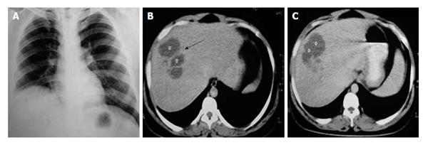
Figure 1 Computed tomography.
A: Chest radiograph shows elevation of right hemi-diaphragm; B: Contrast enhanced computed tomography of upper abdomen shows one loculated hypodense lesion (8.5 cm × 7.4 cm) with irregular inner margin noted in the right lobe of liver (black arrow); C: Multiple small hypodense lesions with confluences also seen in posterior part of right lobe.
-
Citation: Pal P, Ray S, Moulick A, Dey S, Jana A, Banerjee K. Liver abscess caused by
Burkholderia pseudomallei in a young man: A case report and review of literature. World J Clin Cases 2014; 2(10): 604-607 - URL: https://www.wjgnet.com/2307-8960/full/v2/i10/604.htm
- DOI: https://dx.doi.org/10.12998/wjcc.v2.i10.604


