Copyright
©The Author(s) 2025.
World J Clin Cases. Mar 26, 2025; 13(9): 99964
Published online Mar 26, 2025. doi: 10.12998/wjcc.v13.i9.99964
Published online Mar 26, 2025. doi: 10.12998/wjcc.v13.i9.99964
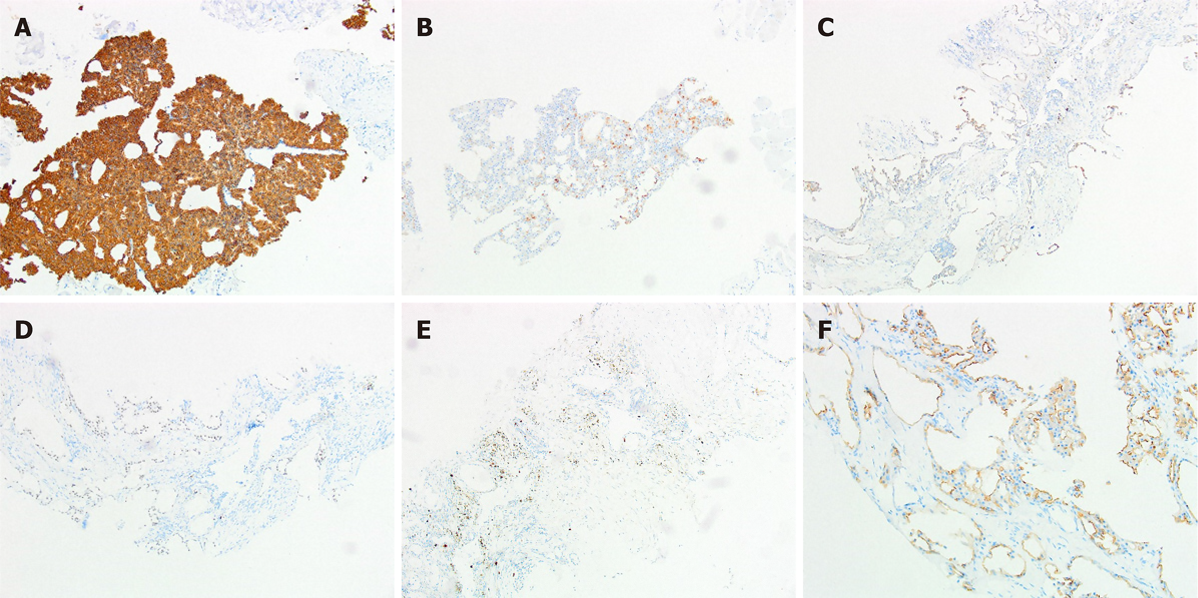
Figure 3 Immunophenotype of this case.
A: Tumor cells were positive for CK pan (× 40); B: Tumor cells were positive for epithelial membrane antigen (× 40); C: Tumor cells were positive for S-100 (× 40); D: Tumor cells nucleus were positive for SOX-10 (× 40); E: Tumor cells were positive in 10% cells for Ki-67 (× 40); F: Tumor cells expressed cell margin membrane DOG-1 (× 40).
- Citation: Sun DQ, Chen CC, Zheng DA, Xing HY, Peng X. Scapular metastasis from acinic cell carcinoma of parotid gland: A case report. World J Clin Cases 2025; 13(9): 99964
- URL: https://www.wjgnet.com/2307-8960/full/v13/i9/99964.htm
- DOI: https://dx.doi.org/10.12998/wjcc.v13.i9.99964


