Copyright
©The Author(s) 2025.
World J Clin Cases. Mar 26, 2025; 13(9): 99421
Published online Mar 26, 2025. doi: 10.12998/wjcc.v13.i9.99421
Published online Mar 26, 2025. doi: 10.12998/wjcc.v13.i9.99421
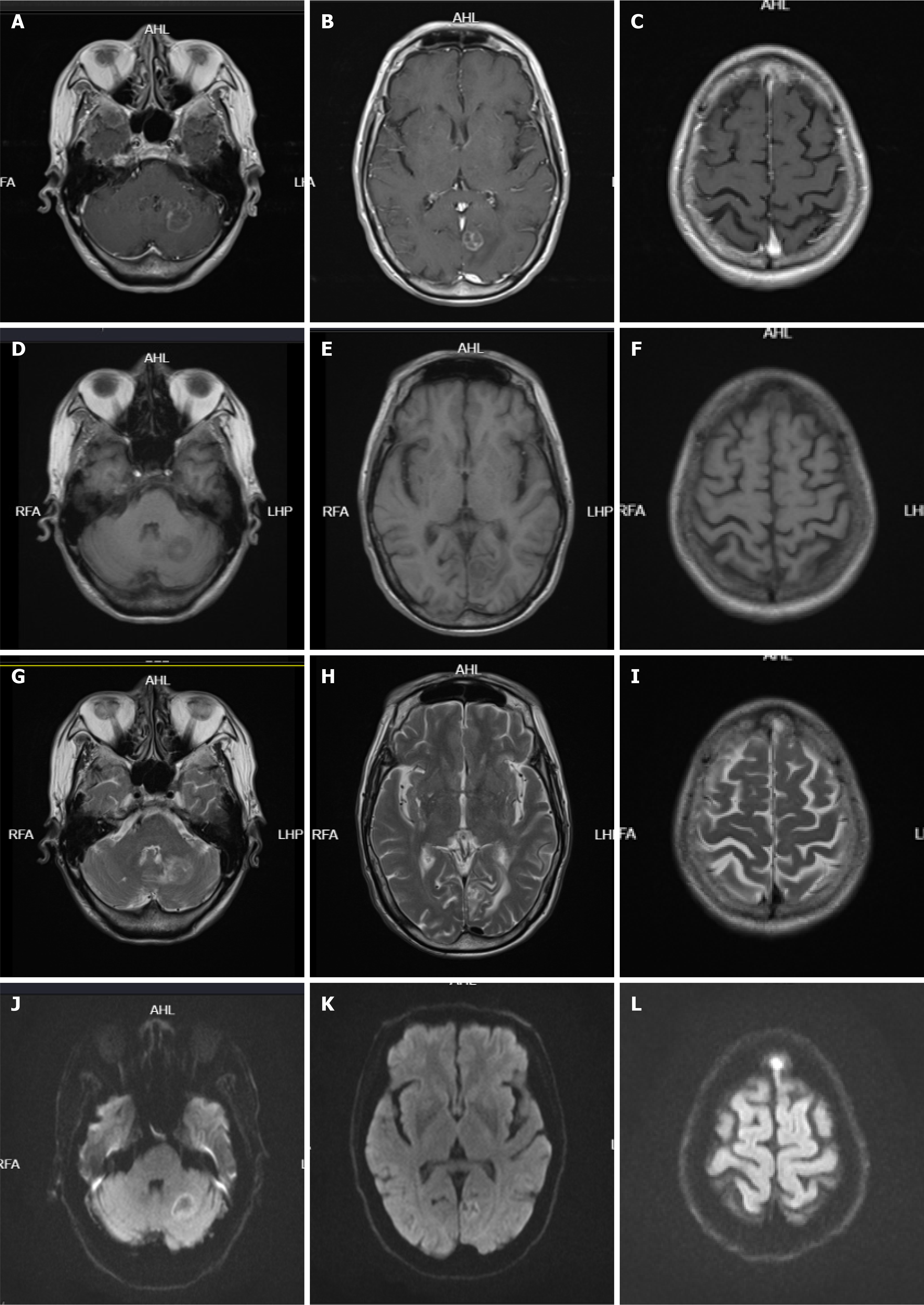
Figure 4 Head magnetic resonance scan and enhancement of Case 1.
A–C: T1-weighted image; D–F: T1 fluid attenuated inversion recovery; G–I: T2-weighted image; J–L: Diffusion-weighted imaging (DWI). The left cerebellar hemisphere and left occipital lobe showed slightly longer circular T1 and slightly longer T2 signals, and the diffusion on DWI was limited. Patchy high signals were seen in the left frontal bone on DWI, with enhanced and visible intensification, about 22 mm × 14 mm in size; patchy abnormal intensification was seen in the right occipital lobe.
- Citation: Luo M, Lu XX, Meng DY, Hu J. Small cell lung cancer with peripheral neuropathy as the first symptom: Two case reports. World J Clin Cases 2025; 13(9): 99421
- URL: https://www.wjgnet.com/2307-8960/full/v13/i9/99421.htm
- DOI: https://dx.doi.org/10.12998/wjcc.v13.i9.99421


