Copyright
©The Author(s) 2025.
World J Clin Cases. Mar 26, 2025; 13(9): 99421
Published online Mar 26, 2025. doi: 10.12998/wjcc.v13.i9.99421
Published online Mar 26, 2025. doi: 10.12998/wjcc.v13.i9.99421
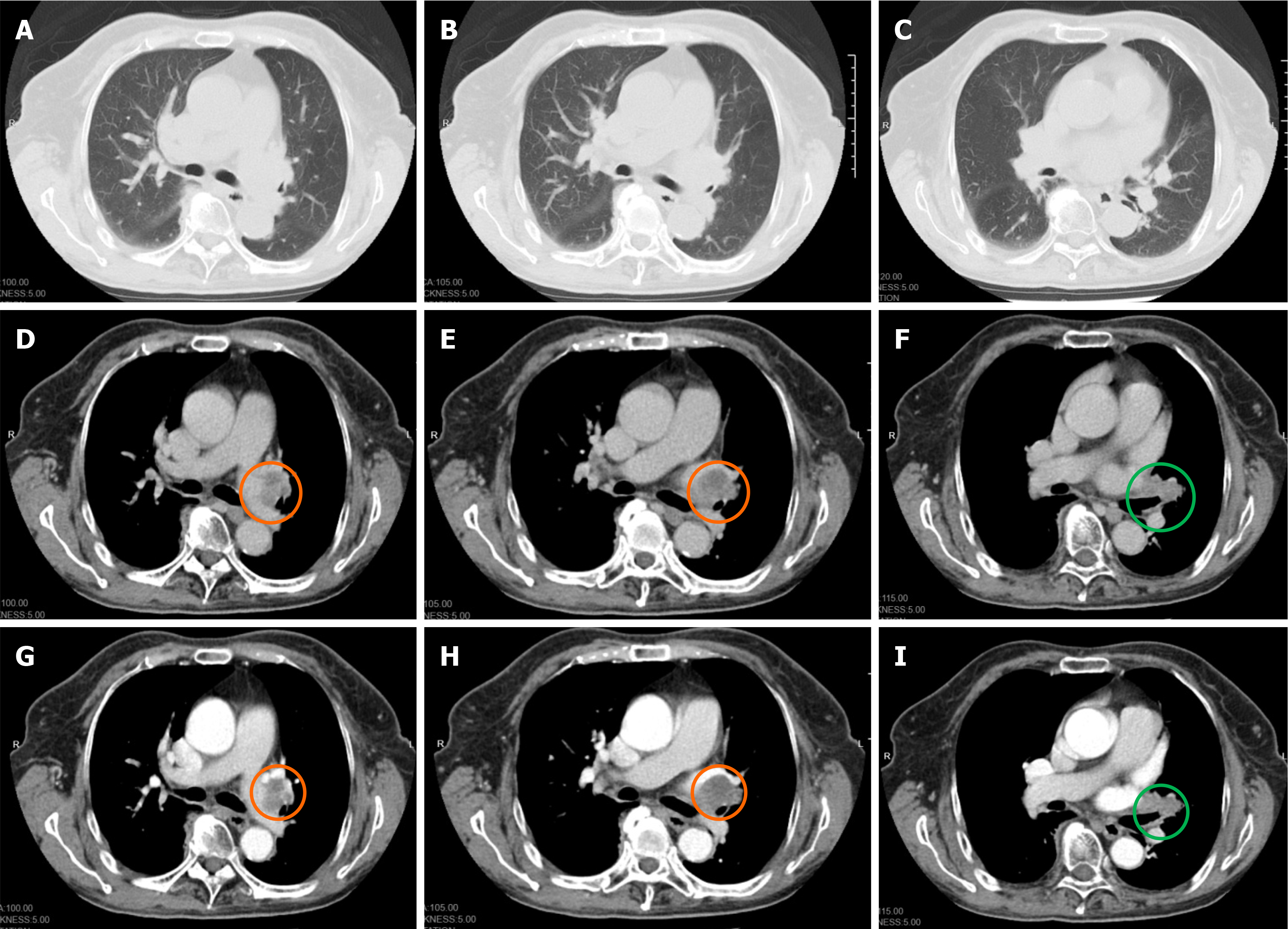
Figure 2 Enhanced lung computed tomography of Case 1.
A–C: Lung view; D–F: Soft tissue view; G–I: Enhanced soft tissue. An irregular soft tissue density shadow with a size of 33 mm × 24 mm was observed at the hilus of the left lung, which showed mild enhancement after enhancement. The bronchial stenosis of the upper lobe of the left lung and pressure of the left upper pulmonary vein were observed. Lymph nodes in the left hilar of the lung were enlarged and enhanced after enhancement. Enlarged lymph nodes are marked in orange and lung cancer is marked in green.
- Citation: Luo M, Lu XX, Meng DY, Hu J. Small cell lung cancer with peripheral neuropathy as the first symptom: Two case reports. World J Clin Cases 2025; 13(9): 99421
- URL: https://www.wjgnet.com/2307-8960/full/v13/i9/99421.htm
- DOI: https://dx.doi.org/10.12998/wjcc.v13.i9.99421


