Copyright
©The Author(s) 2025.
World J Clin Cases. Feb 16, 2025; 13(5): 97629
Published online Feb 16, 2025. doi: 10.12998/wjcc.v13.i5.97629
Published online Feb 16, 2025. doi: 10.12998/wjcc.v13.i5.97629
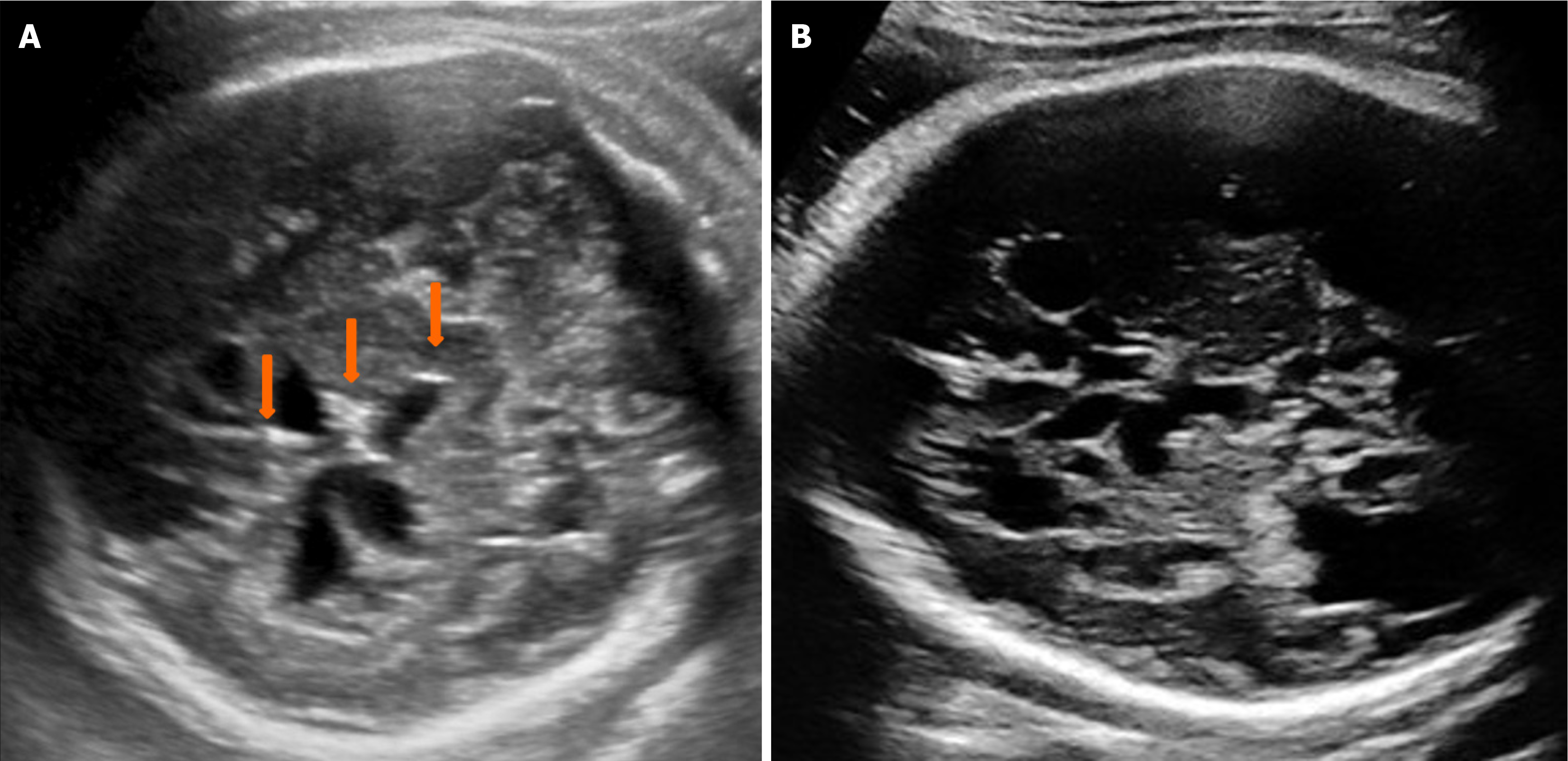
Figure 2 Axial view of the fetal head in the third trimester.
A: The transverse view of the fetal head at 35 weeks of gestation demonstrated midline and intraventricular calcifications (orange arrow); B: At 38 weeks of gestation, the transverse view exhibited multiple cysts, ventriculomegaly, and a smooth brain surface (lissencephaly).
- Citation: Chen XL, Zhang LQ, Bai LL. Ultrasound features of congenital cytomegalovirus infection in the first trimester: A case report. World J Clin Cases 2025; 13(5): 97629
- URL: https://www.wjgnet.com/2307-8960/full/v13/i5/97629.htm
- DOI: https://dx.doi.org/10.12998/wjcc.v13.i5.97629


