Copyright
©The Author(s) 2025.
World J Clin Cases. Jan 26, 2025; 13(3): 99558
Published online Jan 26, 2025. doi: 10.12998/wjcc.v13.i3.99558
Published online Jan 26, 2025. doi: 10.12998/wjcc.v13.i3.99558
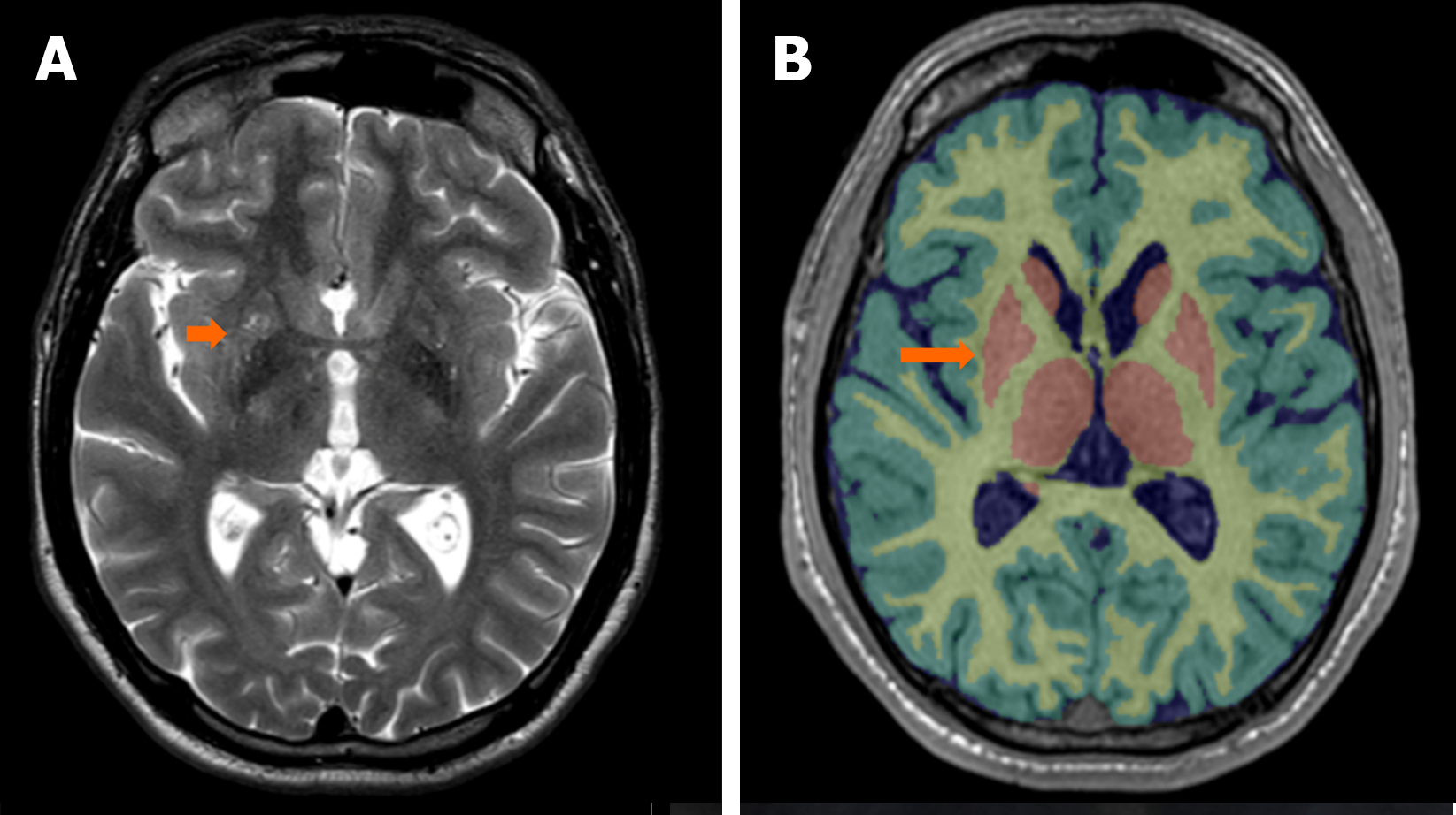
Figure 4 Encephalic atrophic changes in magnetic resonance imaging of Case 1.
A: T2-turbo spin-echo magnetic resonance imaging scan with an axial section view at the level of the anterior commissure. Findings are consistent with multiple diffuse T2 hyperintensities in the lentiform and caudate nuclei. The orange arrow indicates the right lenticular nucleus; B: A magnetic resonance imaging with automated analysis of T1-3D sequence shows pathological findings in the right lentiform nucleus (orange arrow), a part of the basal nuclei. The basal nuclei presented a reduced volume corresponding to less than the 1st percentile for age and sex in our patient.
- Citation: Carrera E, Alvarado J, Astudillo M, Pillajo G. Wilson's disease in two siblings from Ecuador: Two case reports. World J Clin Cases 2025; 13(3): 99558
- URL: https://www.wjgnet.com/2307-8960/full/v13/i3/99558.htm
- DOI: https://dx.doi.org/10.12998/wjcc.v13.i3.99558


