Copyright
©The Author(s) 2025.
World J Clin Cases. Jan 26, 2025; 13(3): 98104
Published online Jan 26, 2025. doi: 10.12998/wjcc.v13.i3.98104
Published online Jan 26, 2025. doi: 10.12998/wjcc.v13.i3.98104
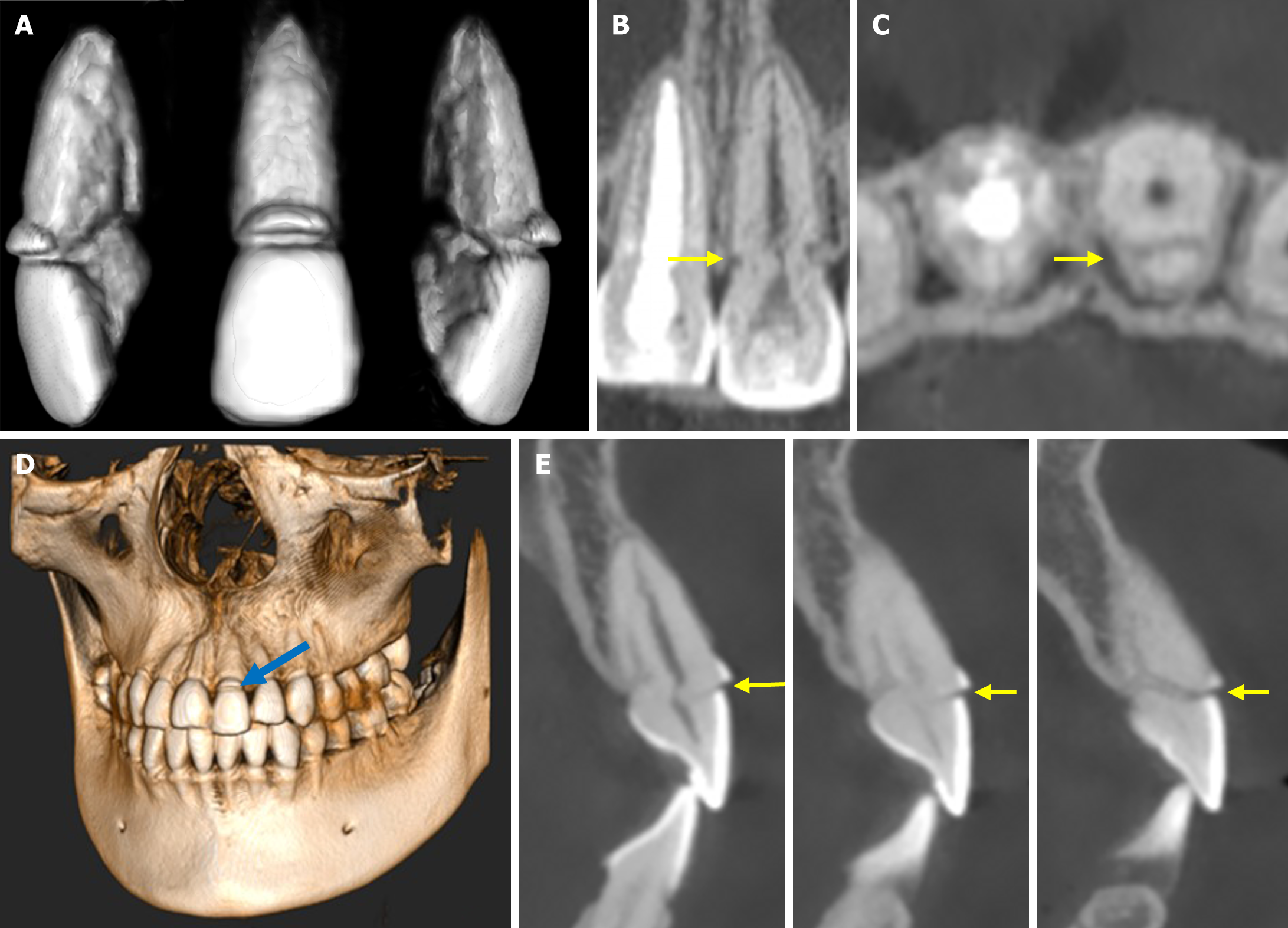
Figure 4 Cone-beam computed tomography views of the fractured central incisor after 8 years.
Favorable healing occurred not only in the crown-root fracture but also in the additional root fracture. A: The three-dimensional (3D) reconstruction highlights the characteristic fracture patterns. The additional fracture starts at the crestal position and extends vertically within the root and backward in an apical direction; B: Cone-beam computed tomography (CBCT) sagittal image showing hard tissue deposition between the fractured surfaces and bulging into the root canal space (arrow); C: CBCT axial slice showing that the additional root fracture line has been obliterated by calcified tissue (arrow); D: The 3D reconstruction indicates that the narrow enamel gutter was above the level of the alveolar bone crest (blue arrow); E: CBCT sections in the coronal plane show that multiple fracture lines have been obliterated by calcified tissue, the arrow indicating the narrow unfilled enamel gutter.
- Citation: Li N, Ren YY, Tang Y, Yang Q, Meng TT, Li S, Zhang J. Pulp health and calcific healing of a complicated crown–root fracture with additional root fracture in a maxillary incisor: A case report. World J Clin Cases 2025; 13(3): 98104
- URL: https://www.wjgnet.com/2307-8960/full/v13/i3/98104.htm
- DOI: https://dx.doi.org/10.12998/wjcc.v13.i3.98104


