Copyright
©The Author(s) 2025.
World J Clin Cases. Jan 26, 2025; 13(3): 98104
Published online Jan 26, 2025. doi: 10.12998/wjcc.v13.i3.98104
Published online Jan 26, 2025. doi: 10.12998/wjcc.v13.i3.98104
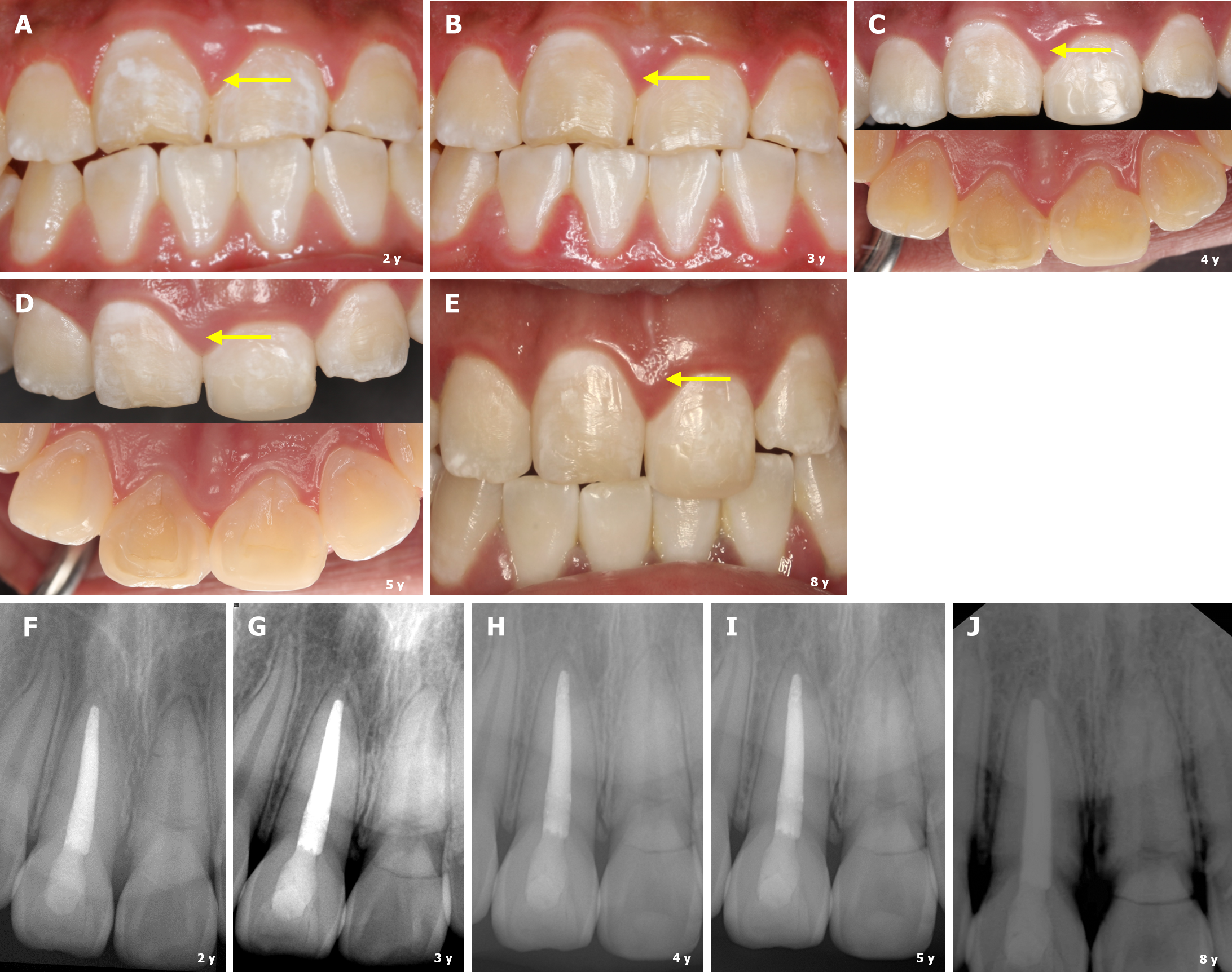
Figure 3 Follow-up clinical views and periapical radiographs from 2015 to 2022, indicated the healed crown-root fracture and a stable prognosis.
A-E: Labial and palatal views of the two involved teeth, which have remained functional and symptomless after removal of the splint (yellow arrow indicating the localized gingival overgrowth); F-J: Periapical radiograph of the teeth showing the progressive increase in calcification within the fracture lines, with no obvious periradicular pathosis.
- Citation: Li N, Ren YY, Tang Y, Yang Q, Meng TT, Li S, Zhang J. Pulp health and calcific healing of a complicated crown–root fracture with additional root fracture in a maxillary incisor: A case report. World J Clin Cases 2025; 13(3): 98104
- URL: https://www.wjgnet.com/2307-8960/full/v13/i3/98104.htm
- DOI: https://dx.doi.org/10.12998/wjcc.v13.i3.98104


