Copyright
©The Author(s) 2025.
World J Clin Cases. Jul 26, 2025; 13(21): 105066
Published online Jul 26, 2025. doi: 10.12998/wjcc.v13.i21.105066
Published online Jul 26, 2025. doi: 10.12998/wjcc.v13.i21.105066
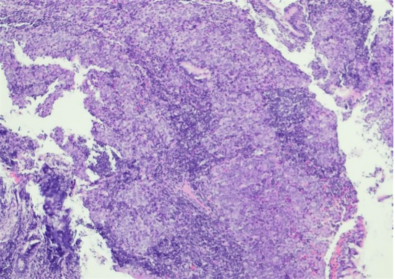
Figure 3 Pathology.
Two grayish-white to yellowish rice-grain-sized masses were obtained from the nasopharynx of the patient. Immunohistochemical analysis revealed the following results: Cytokeratin (+), tumor Protein p53 (wild-type), tumor Protein p63(+), marker of Proliferation Ki-67 (30%+), tumor Necrosis Factor Receptor Superfamily Member 8 (scattered+), ΔNp63 Isoform of tumor Protein p63 (P40) (+), Cytokeratin 7 (-), and Cytokeratin 5/6 (+). Hematoxylin and eosin staining; magnification, ×100.
- Citation: Zhou XY, Jiang YJ, Guo XM, Han DH, Liu Y, Qiao Q. Application of circulating tumor DNA liquid biopsy in nasopharyngeal carcinoma: A case report and review of literature. World J Clin Cases 2025; 13(21): 105066
- URL: https://www.wjgnet.com/2307-8960/full/v13/i21/105066.htm
- DOI: https://dx.doi.org/10.12998/wjcc.v13.i21.105066


