Copyright
©The Author(s) 2025.
World J Clin Cases. Jul 26, 2025; 13(21): 104062
Published online Jul 26, 2025. doi: 10.12998/wjcc.v13.i21.104062
Published online Jul 26, 2025. doi: 10.12998/wjcc.v13.i21.104062
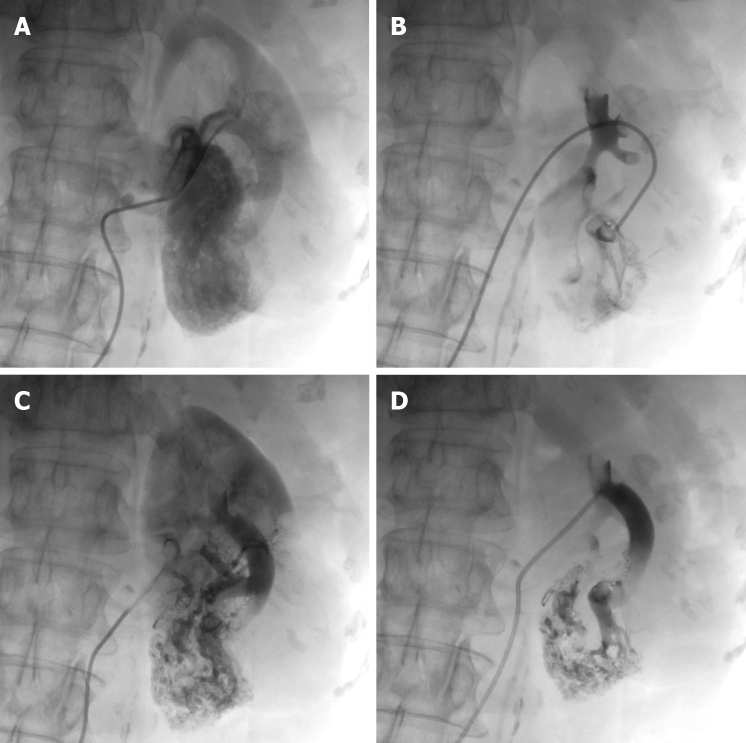
Figure 6 Digital subtraction angiography examination of the left renal artery.
A and B: Rapid visualization of the left renal vein. An arteriovenous fistula formation was observed in 4 branches of the left renal artery, contrast extravasation was observed, along with a large lamellar vascular malformation in the lower pole of the left kidney; C and D: The diseased artery was embolized with a spring coil. Postembolization review revealed complete embolization of the diseased artery, closure of the arteriovenous fistula, and no early visualization of the left renal vein.
- Citation: Lv SP, Qin LL, Mou H, Huang T, Wang KQ. Multimodal imaging techniques for the diagnosis of congenital left renal arteriovenous fistula: A case report. World J Clin Cases 2025; 13(21): 104062
- URL: https://www.wjgnet.com/2307-8960/full/v13/i21/104062.htm
- DOI: https://dx.doi.org/10.12998/wjcc.v13.i21.104062


