Copyright
©The Author(s) 2025.
World J Clin Cases. Jul 26, 2025; 13(21): 104062
Published online Jul 26, 2025. doi: 10.12998/wjcc.v13.i21.104062
Published online Jul 26, 2025. doi: 10.12998/wjcc.v13.i21.104062
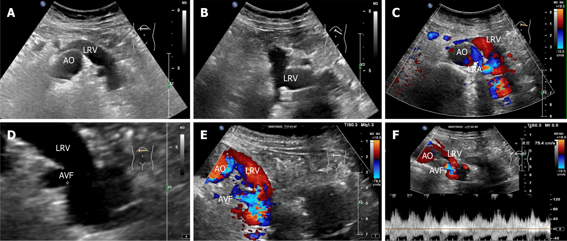
Figure 3 Renal vascular ultrasonography.
A and B: Two-dimensional image showing that the left renal vein is widened and uneven in thickness; C: Color doppler flow imaging reveals a brighter florid blood flow signal within the left renal vein, with the blood-supplying artery originating from the adjacent left renal artery; D: Magnified two-dimensional image showing a 0.5 cm wide fistula between the left renal vein and the left renal artery; E: Color doppler flow imaging reveals a bright florid blood flow signal in the left renal vein near the fistula; F: Spectral doppler reveals spectral arterialization of the left renal vein with a non-smooth envelope and burr-like changes. AO: Abdominal aorta; LRV: Left renal vein; LRA: Left renal artery; AVF: Arteriovenous fistula.
- Citation: Lv SP, Qin LL, Mou H, Huang T, Wang KQ. Multimodal imaging techniques for the diagnosis of congenital left renal arteriovenous fistula: A case report. World J Clin Cases 2025; 13(21): 104062
- URL: https://www.wjgnet.com/2307-8960/full/v13/i21/104062.htm
- DOI: https://dx.doi.org/10.12998/wjcc.v13.i21.104062


