Copyright
©The Author(s) 2025.
World J Clin Cases. Jul 26, 2025; 13(21): 103105
Published online Jul 26, 2025. doi: 10.12998/wjcc.v13.i21.103105
Published online Jul 26, 2025. doi: 10.12998/wjcc.v13.i21.103105
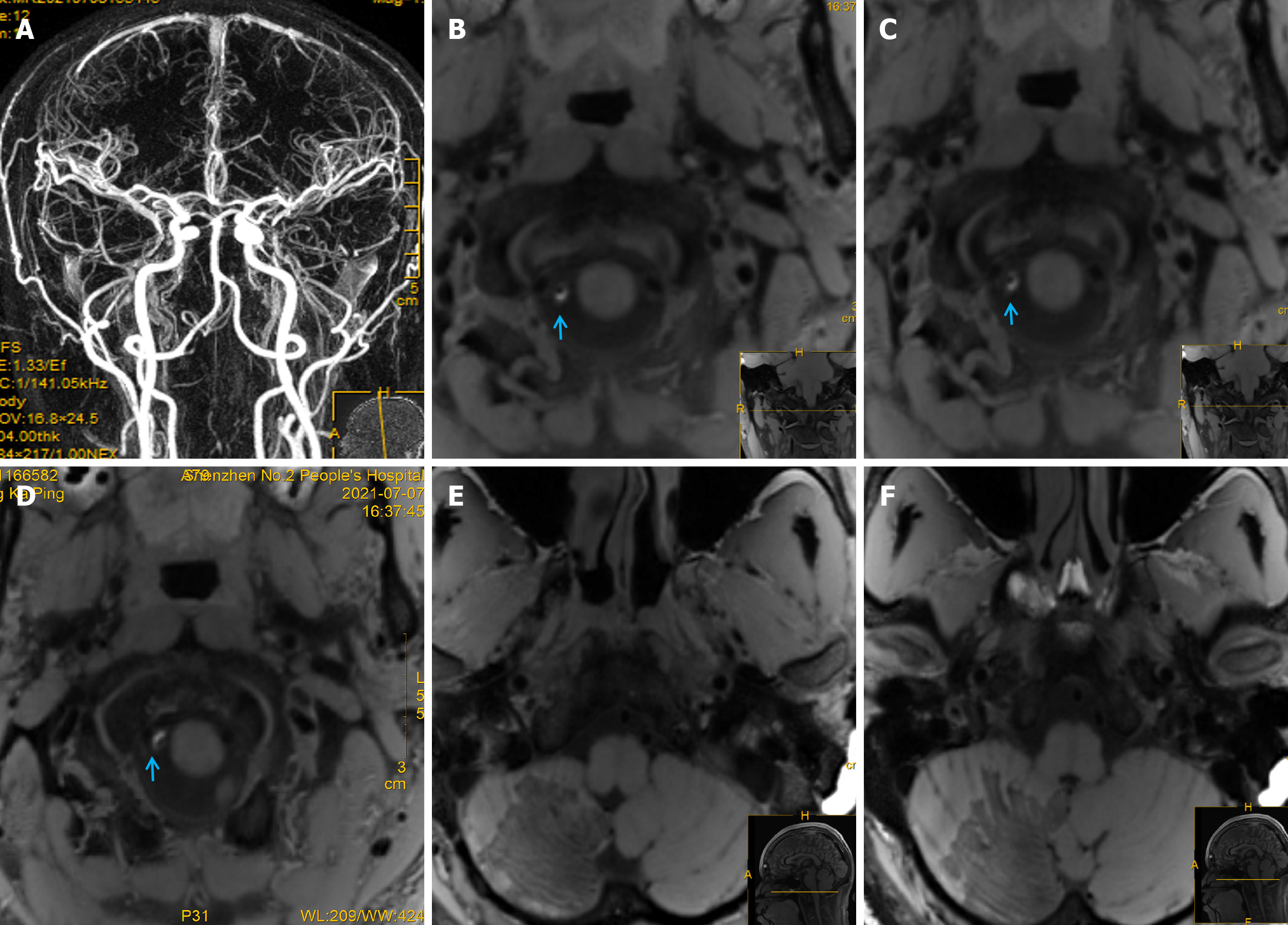
Figure 2 Findings on magnetic resonance angiography, high-resolution vessel-wall magnetic resonance imaging, and follow-up imaging findings.
A: Brain magnetic resonance angiography (MRA) shows no obvious abnormalities; B-D: High-resolution vessel-wall magnetic resonance imaging (HR-VW MRI) reveals a crescent-shaped enhanced lesion in the vascular wall of the posterior inferior cerebellar artery (arrows); E and F: 1.5-month follow-up HR-VW MRI demonstrating complete disappearance of the lesion.
- Citation: Huang XM, Liao YQ, Cao LM. Massive cerebellar infarction caused by spontaneously isolated posterior inferior cerebellar artery dissection: A case report. World J Clin Cases 2025; 13(21): 103105
- URL: https://www.wjgnet.com/2307-8960/full/v13/i21/103105.htm
- DOI: https://dx.doi.org/10.12998/wjcc.v13.i21.103105


