Copyright
©The Author(s) 2025.
World J Clin Cases. Jul 16, 2025; 13(20): 102695
Published online Jul 16, 2025. doi: 10.12998/wjcc.v13.i20.102695
Published online Jul 16, 2025. doi: 10.12998/wjcc.v13.i20.102695
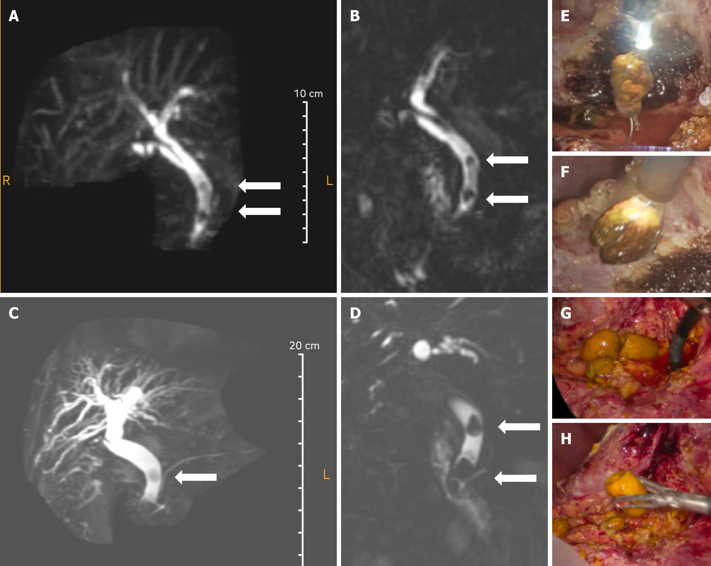
Figure 2 T2 coronal image shows bi-lobar central and peripheral intrahepatic biliary tract dilatation and dilated common bile duct with distal smooth tapering of common bile duct.
White solid arrow: The filling defect.
- Citation: Ren ZH, Gao YY, Lu Q, Yao YM, Wan Y. Rare recurrence of common bile duct calculi post T-tube cholangiography: A case report. World J Clin Cases 2025; 13(20): 102695
- URL: https://www.wjgnet.com/2307-8960/full/v13/i20/102695.htm
- DOI: https://dx.doi.org/10.12998/wjcc.v13.i20.102695


