Copyright
©The Author(s) 2025.
World J Clin Cases. Jul 16, 2025; 13(20): 102279
Published online Jul 16, 2025. doi: 10.12998/wjcc.v13.i20.102279
Published online Jul 16, 2025. doi: 10.12998/wjcc.v13.i20.102279
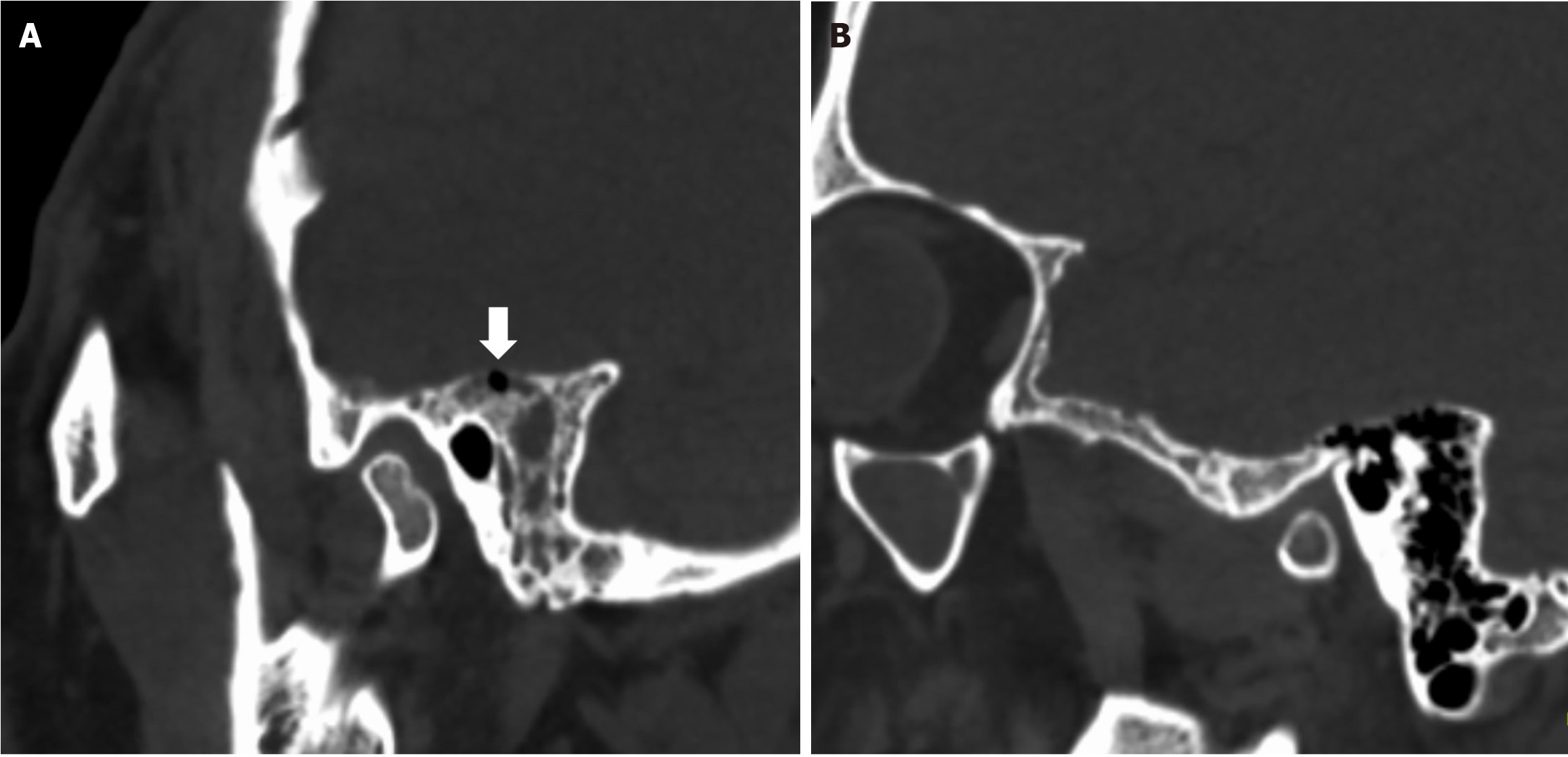
Figure 1 Preoperative high-resolution computed tomography image of the temporal bone.
A: Left temporal bone and mastoid process (affected side). A rough and damaged bony structure of the petrous part of the temporal bone, with fluid accumulation in the left mastoid process, left middle ear, and external ear canal was observed; B: Right temporal bone and mastoid process (healthy side). White arrows indicate suspected bone defect locations.
- Citation: He YS, Zheng Y. Exploratory operation in a patient with spontaneous temporal bone cerebrospinal fluid leaks: A case report. World J Clin Cases 2025; 13(20): 102279
- URL: https://www.wjgnet.com/2307-8960/full/v13/i20/102279.htm
- DOI: https://dx.doi.org/10.12998/wjcc.v13.i20.102279


