Copyright
©The Author(s) 2025.
World J Clin Cases. Jan 16, 2025; 13(2): 96876
Published online Jan 16, 2025. doi: 10.12998/wjcc.v13.i2.96876
Published online Jan 16, 2025. doi: 10.12998/wjcc.v13.i2.96876
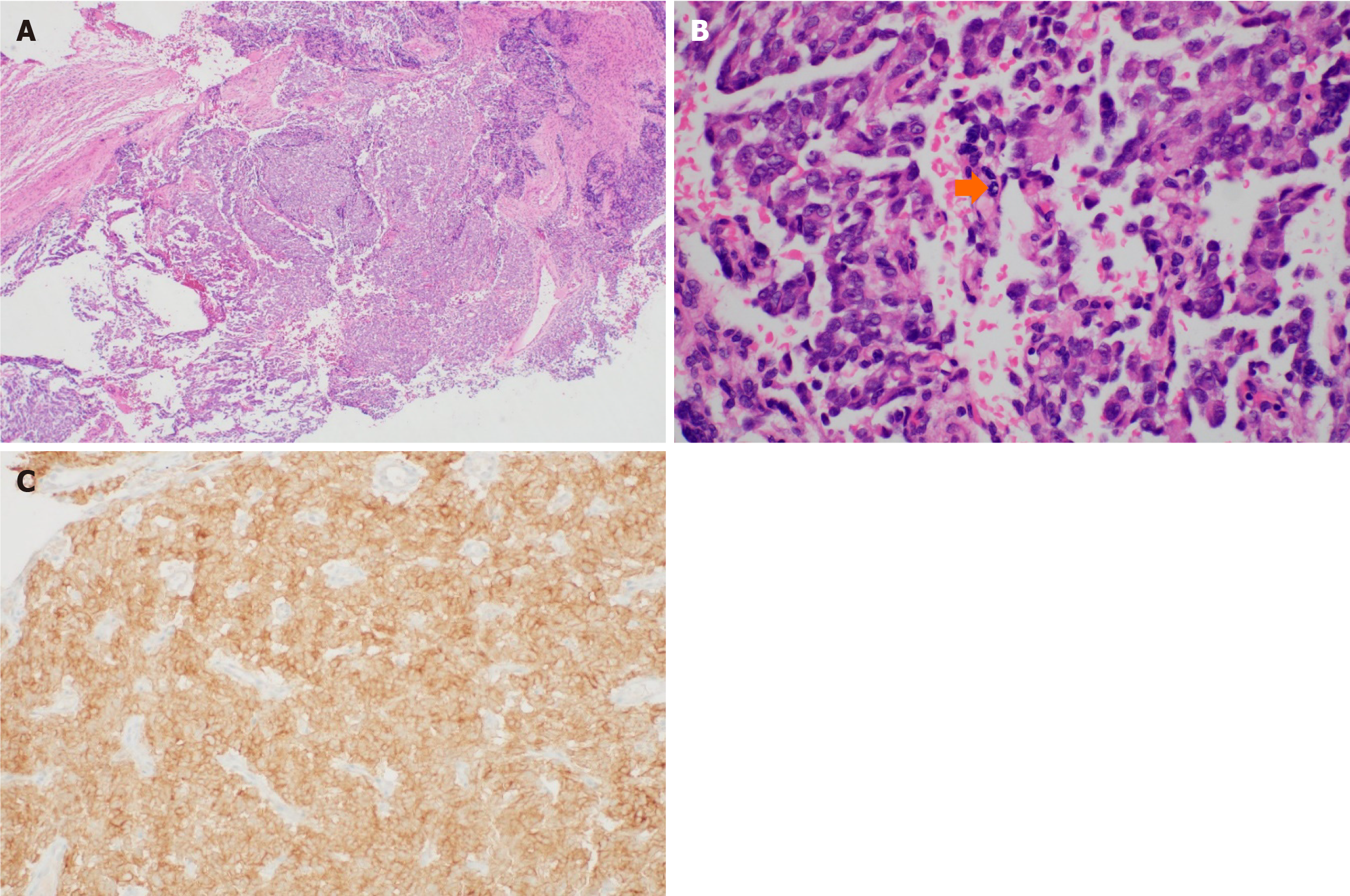
Figure 4 Pan-TRK staining was positive.
A and B: Infiltrating tumor cells in a sheet pattern were observed in cervical biopsy (A) [hematoxylin-eosin (HE) stain, × 40]. Round or oval cells have a vesicular nucleus and distinct nucleoli. Mitotic activities were commonly observed and there were also some necrotic areas (arrow) (B) (HE stain, × 400); C: As in the cytology specimen, pan-TRK staining was positive (× 200).
- Citation: Lee S, Jeon YR, Shin C, Kwon SY, Shin S. Pan-TRK positive uterine sarcoma in immunohistochemistry without neurotrophic tyrosine receptor kinase gene fusions: A case report. World J Clin Cases 2025; 13(2): 96876
- URL: https://www.wjgnet.com/2307-8960/full/v13/i2/96876.htm
- DOI: https://dx.doi.org/10.12998/wjcc.v13.i2.96876


