Copyright
©The Author(s) 2025.
World J Clin Cases. Jan 16, 2025; 13(2): 96876
Published online Jan 16, 2025. doi: 10.12998/wjcc.v13.i2.96876
Published online Jan 16, 2025. doi: 10.12998/wjcc.v13.i2.96876
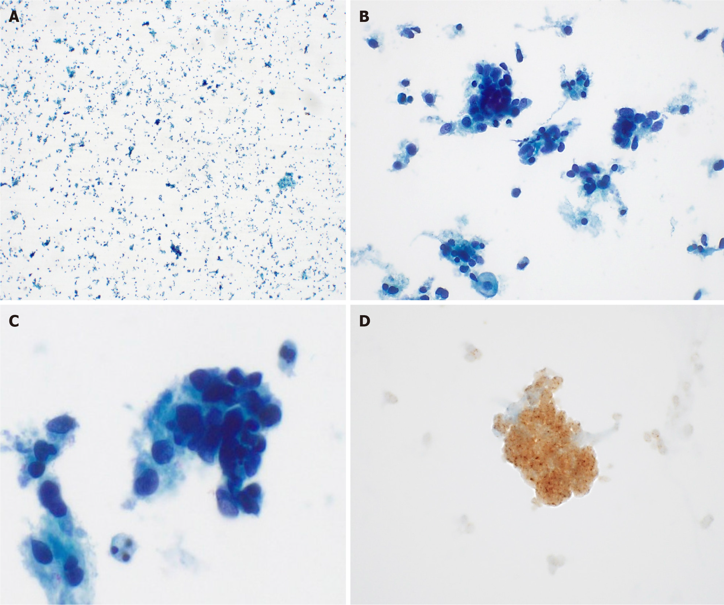
Figure 3 Liquid-based cytology detected ovoid to round cells with hyperchromatic nuclei.
Immunohistochemistry exhibited diffuse strong positivity for pan-TRK. A and B: Low power view of uterine cervix (A) (Papanicoalu stain, × 40) A cluster of atypical cells with clumped chromatin is observed, and some cells with low cohesion are scattered individually (B) (Papanicoalu stain, × 100); C and D: The tumor cells were ovoid to round cells with hyperchromatic nuclei formed loosely cohesive clusters without keratinization (C) (Papanicoalu stain, × 400). Pan-TRK staining showed diffuse strong positivity in cytologic specimen (D) (× 400).
- Citation: Lee S, Jeon YR, Shin C, Kwon SY, Shin S. Pan-TRK positive uterine sarcoma in immunohistochemistry without neurotrophic tyrosine receptor kinase gene fusions: A case report. World J Clin Cases 2025; 13(2): 96876
- URL: https://www.wjgnet.com/2307-8960/full/v13/i2/96876.htm
- DOI: https://dx.doi.org/10.12998/wjcc.v13.i2.96876


