Copyright
©The Author(s) 2025.
World J Clin Cases. Jan 16, 2025; 13(2): 96876
Published online Jan 16, 2025. doi: 10.12998/wjcc.v13.i2.96876
Published online Jan 16, 2025. doi: 10.12998/wjcc.v13.i2.96876
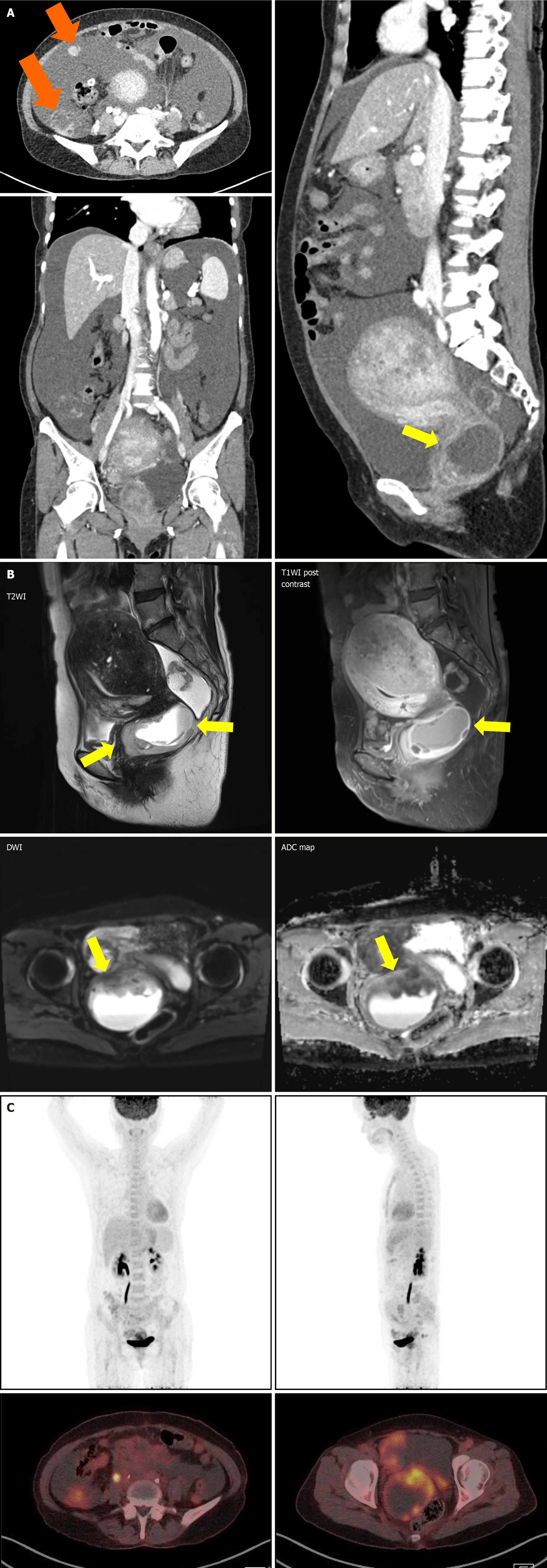
Figure 2 Imaging at diagnosis.
A: Pelvic and chest computed tomography; B: Pelvic magnetic resonance imaging; C: Positron emission tomography-computed tomography. A 5 cm mass with necrosis in the uterine cervix is observed (arrows, yellow), along with multiple peritoneal seeding in the right paracolic gutter (arrows, orange) and a large amount of ascites. T2WI: T2 weighted image; T1WI: T1 weighted image; DWI: Diffusion weighted image; ADC: Apparent diffusion coefficient.
- Citation: Lee S, Jeon YR, Shin C, Kwon SY, Shin S. Pan-TRK positive uterine sarcoma in immunohistochemistry without neurotrophic tyrosine receptor kinase gene fusions: A case report. World J Clin Cases 2025; 13(2): 96876
- URL: https://www.wjgnet.com/2307-8960/full/v13/i2/96876.htm
- DOI: https://dx.doi.org/10.12998/wjcc.v13.i2.96876


