Copyright
©The Author(s) 2025.
World J Clin Cases. Jul 6, 2025; 13(19): 103946
Published online Jul 6, 2025. doi: 10.12998/wjcc.v13.i19.103946
Published online Jul 6, 2025. doi: 10.12998/wjcc.v13.i19.103946
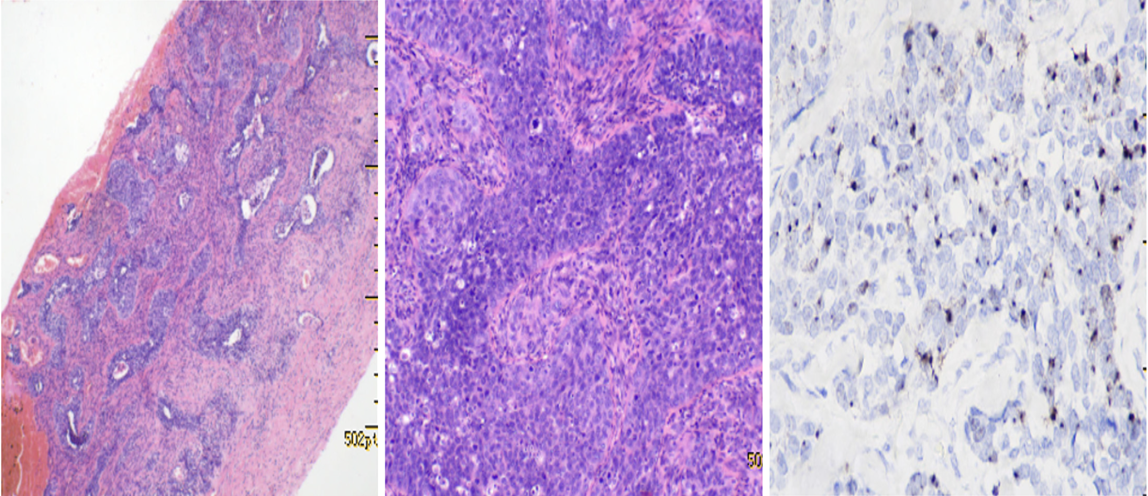
Figure 3 Pathological pictures of the patient.
Histology of serous carcinoma of the meningeal metastases shows endometrioid gland lined by columnar cells with eosinophilic cytoplasm and pseudostratified nuclei (hematoxylin and eosin × 400, left). Multi-subtype in situ hybridization showing spotty and patchy positive signals in the cytoplasm of tumor cells (right).
- Citation: Huang HQ, Gong FM, Sun CT, Xuan Y, Li L. Brain and scalp metastasis of cervical cancer in a patient with human immunodeficiency virus infection: A case report. World J Clin Cases 2025; 13(19): 103946
- URL: https://www.wjgnet.com/2307-8960/full/v13/i19/103946.htm
- DOI: https://dx.doi.org/10.12998/wjcc.v13.i19.103946


