Copyright
©The Author(s) 2025.
World J Clin Cases. Jul 6, 2025; 13(19): 103946
Published online Jul 6, 2025. doi: 10.12998/wjcc.v13.i19.103946
Published online Jul 6, 2025. doi: 10.12998/wjcc.v13.i19.103946
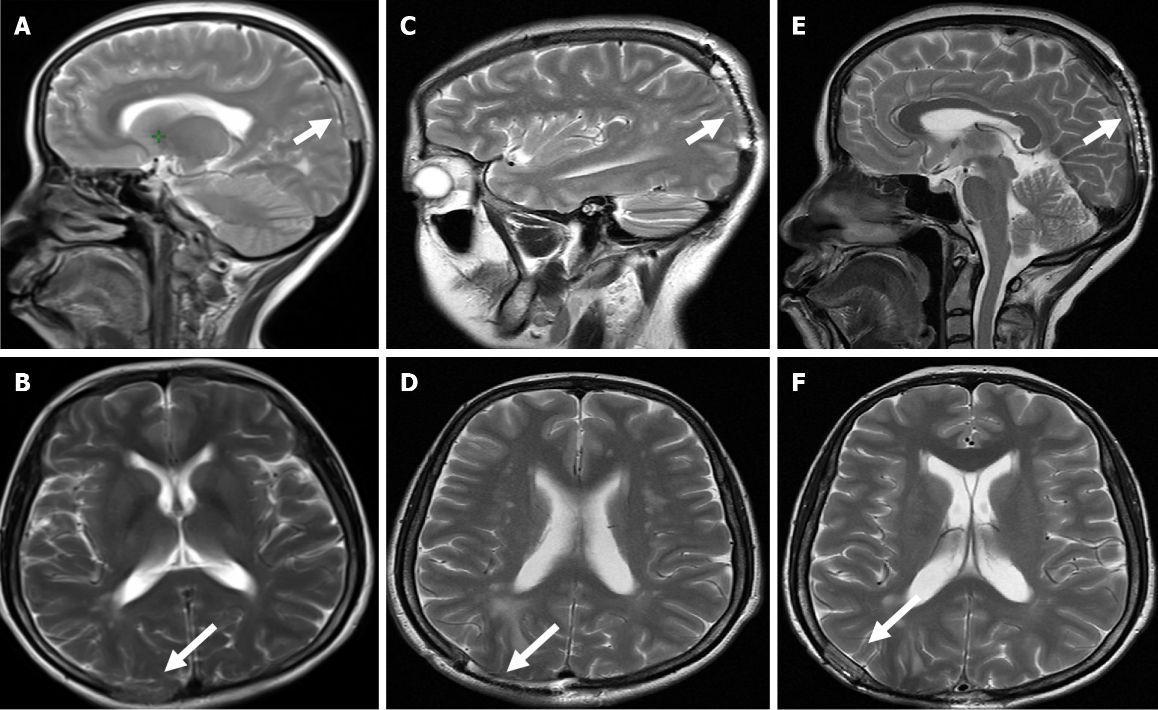
Figure 2 Images of brain magnetic resonance imaging.
A and B: Patchy high signal shadow was observed between the right parietal bone and adjacent soft tissues, with limited diffusion (white arrows); C and D: Brain magnetic resonance imaging before radiotherapy showed postoperative changes, and a cranial bone flap can be seen; E and F: Brain magnetic resonance imaging images obtained 6 months after the treatment (white arrows).
- Citation: Huang HQ, Gong FM, Sun CT, Xuan Y, Li L. Brain and scalp metastasis of cervical cancer in a patient with human immunodeficiency virus infection: A case report. World J Clin Cases 2025; 13(19): 103946
- URL: https://www.wjgnet.com/2307-8960/full/v13/i19/103946.htm
- DOI: https://dx.doi.org/10.12998/wjcc.v13.i19.103946


