Copyright
©The Author(s) 2025.
World J Clin Cases. Jul 6, 2025; 13(19): 102212
Published online Jul 6, 2025. doi: 10.12998/wjcc.v13.i19.102212
Published online Jul 6, 2025. doi: 10.12998/wjcc.v13.i19.102212
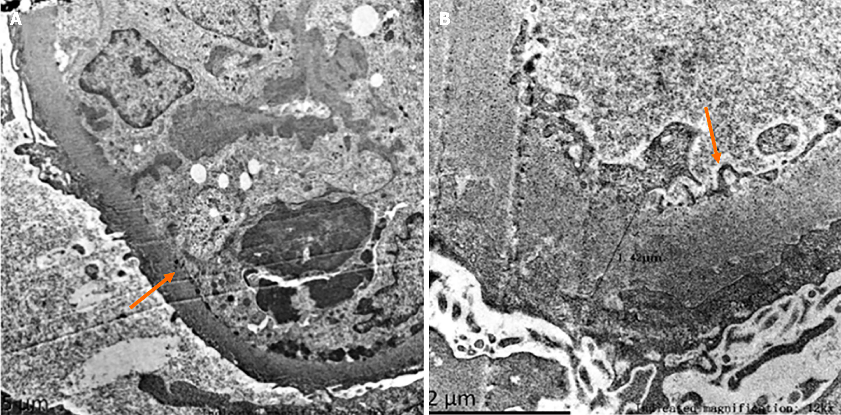
Figure 3 Electron microscopic picture of renal pathology.
Electron microscopy reveals mild to moderate glomerular mesangial hyperplasia accompanied by segmental endothelial cell proliferation and neutrophil infiltration. Multiple immune complexes are identified, along with diffuse basement membrane thickening, the formation of small pegs, extensive fusion of pedicles, and the characteristic double-tracking and layering pattern. Multifocal renal tubular atrophy is observed, along with edema, vacuolar degeneration in some tubular epithelial cells, interstitial edema, focal fibrous tissue hyperplasia, and scattered inflammatory cell infiltration. A: Diffuse basement membrane thickening (orange arrow); B: The formation of small pegs (orange arrow).
- Citation: Li MR, Li LY, Tang J, Sun J. Chronic hepatitis B triggering antineutrophil cytoplasmic antibody-associated vasculitis complicated by glomerulonephritis: A case report. World J Clin Cases 2025; 13(19): 102212
- URL: https://www.wjgnet.com/2307-8960/full/v13/i19/102212.htm
- DOI: https://dx.doi.org/10.12998/wjcc.v13.i19.102212


