Copyright
©The Author(s) 2025.
World J Clin Cases. Jul 6, 2025; 13(19): 102212
Published online Jul 6, 2025. doi: 10.12998/wjcc.v13.i19.102212
Published online Jul 6, 2025. doi: 10.12998/wjcc.v13.i19.102212
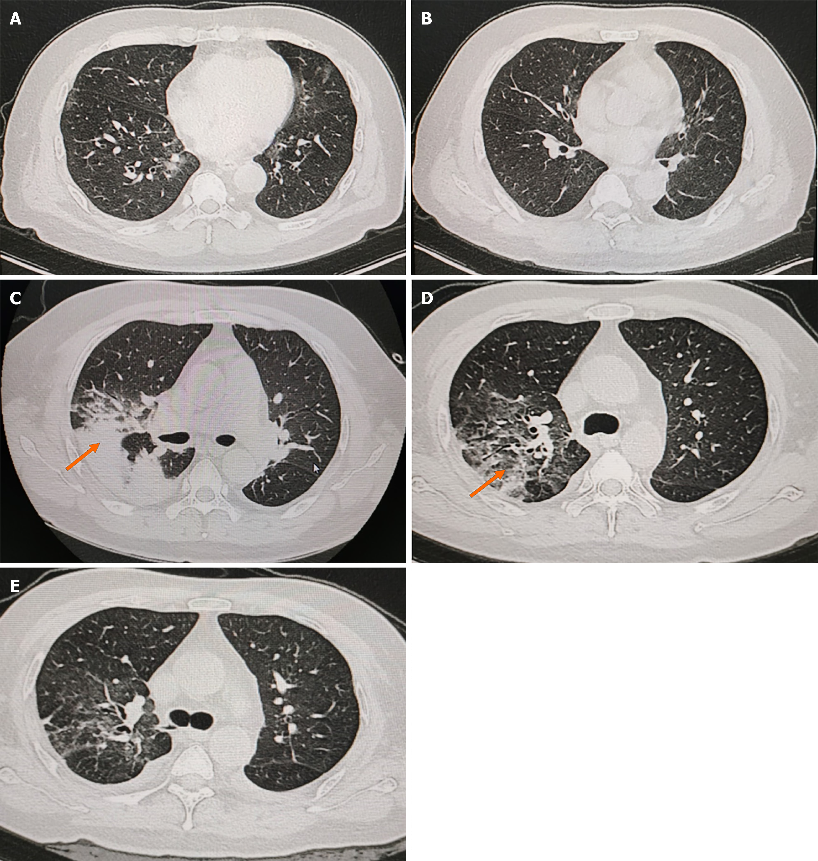
Figure 1 Computed tomography imaging images at the time of initial hospitalization and disease progression.
A and B: High-resolution computed tomography (CT) images to consider viral infections in both lungs; C: Pulmonary artery imaging on March 23, 2023, reveals multiple inflammations in both lungs are significantly more advanced than before. CT angiography imaging of the pulmonary arteries reveals no significant abnormalities. The orange arrow shows prominent solid exudative changes in the right upper lung; D: Multiple inflammations in both lungs are significantly more resorbed than before; E: High-resolution CT of the lungs, performed on April 13, 2023, demonstrates partial resolution of the lesions in the upper and dorsal segments of the right upper and lower lobes, along with a small amount of bilateral pleural fluid effusion. Moreover, partial distension of the adjacent lower lungs is observed.
- Citation: Li MR, Li LY, Tang J, Sun J. Chronic hepatitis B triggering antineutrophil cytoplasmic antibody-associated vasculitis complicated by glomerulonephritis: A case report. World J Clin Cases 2025; 13(19): 102212
- URL: https://www.wjgnet.com/2307-8960/full/v13/i19/102212.htm
- DOI: https://dx.doi.org/10.12998/wjcc.v13.i19.102212


