Copyright
©The Author(s) 2025.
World J Clin Cases. Jun 16, 2025; 13(17): 101593
Published online Jun 16, 2025. doi: 10.12998/wjcc.v13.i17.101593
Published online Jun 16, 2025. doi: 10.12998/wjcc.v13.i17.101593
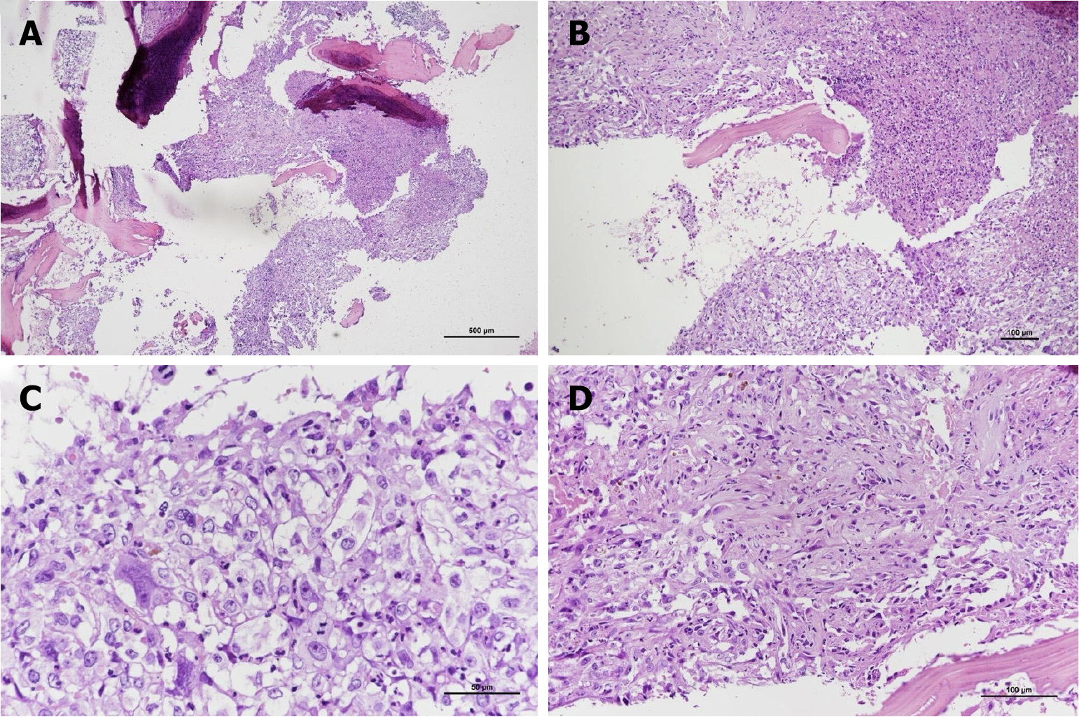
Figure 3 Histopathological examination of the tumor.
Proliferation of cells exhibiting epithelioid morphology, prominent nucleoli and abundant eosinophilic cytoplasm. Tumor necrosis is present. A: Hematoxylin-eosin (HE), 40 ×; B: HE, 100 ×; C: High-power view demonstrates pleomorphic epithelioid cells, intermixed with acute inflammation. Mitotic cells are present (HE, 400 ×); D: Anastomotic vascular channels lined by epithelioid endothelial cells (HE, 200 ×).
- Citation: Nan YH, Chiu CD, Chen WL, Chen LC, Chen CC, Cho DY, Guo JH. Epithelioid angiosarcoma of the cervical spine: A case report. World J Clin Cases 2025; 13(17): 101593
- URL: https://www.wjgnet.com/2307-8960/full/v13/i17/101593.htm
- DOI: https://dx.doi.org/10.12998/wjcc.v13.i17.101593


