Copyright
©The Author(s) 2025.
World J Clin Cases. Jun 16, 2025; 13(17): 101593
Published online Jun 16, 2025. doi: 10.12998/wjcc.v13.i17.101593
Published online Jun 16, 2025. doi: 10.12998/wjcc.v13.i17.101593
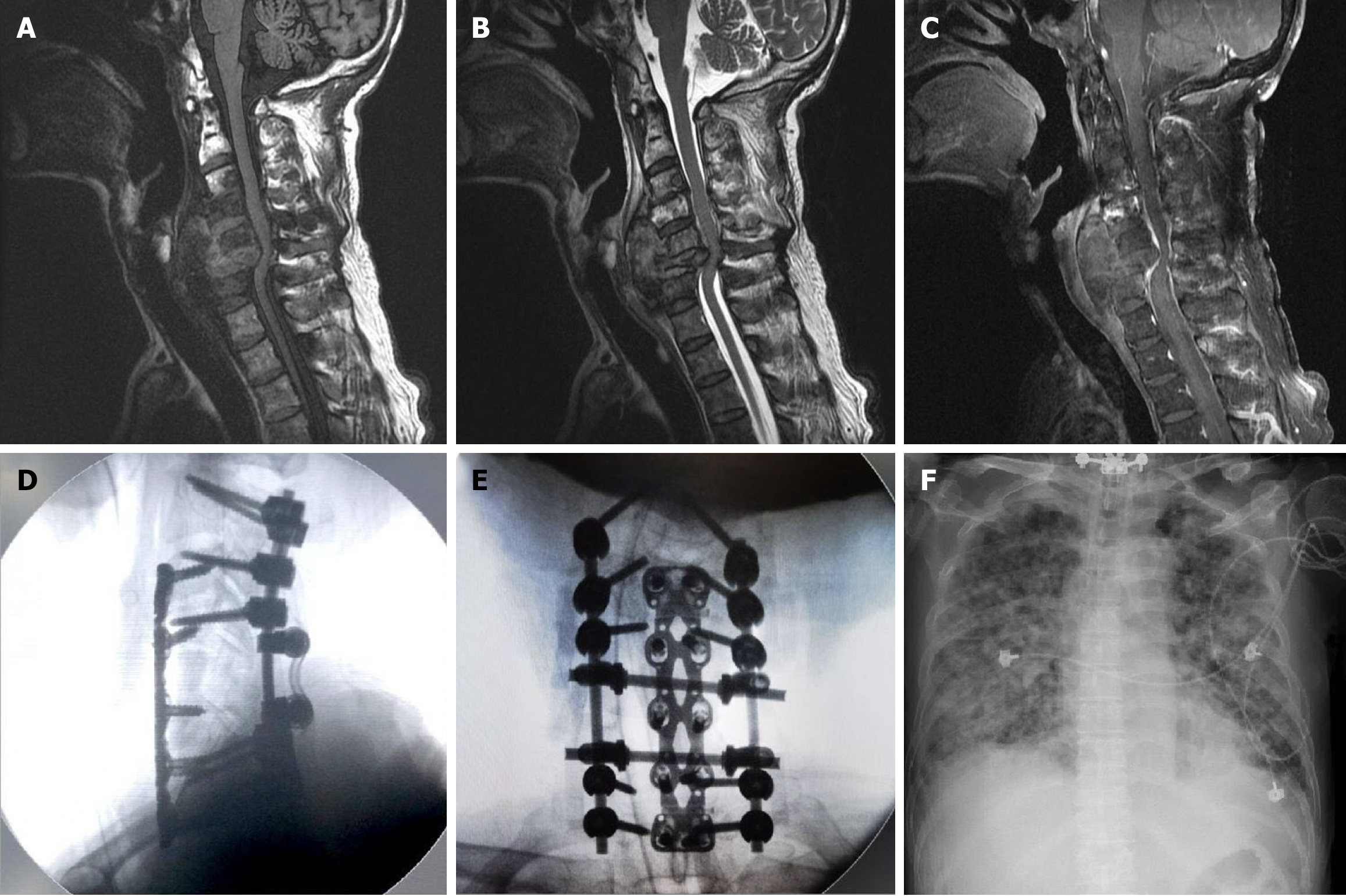
Figure 2 Pre-operative cervical spine magnetic resonance imaging, intra-operative C-arm radiograph, and post-operative chest radiograph.
A: Spinal magnetic resonance images demonstrate the C6 pathologic fracture on T1-weighted images; B: With central necrosis and mixed some fluid and hematoma on T2-weighted images; C: Poor and heterogenous enhancement on T1-weighted images with gadolinium administration; D: The C-arm radiograph images show C2/3/4/7/T1 transpedicular screw and C5-6 corpectomy with tibia bone graft fusion and plate fixation in lateral view; E: The C-arm radiograph images show C2/3/4/7/T1 transpedicular screw and C5-6 corpectomy with tibia bone graft fusion and plate fixation in coronal view; F: Rapid progression of acute respiratory distress syndrome 3 weeks after the second surgery.
- Citation: Nan YH, Chiu CD, Chen WL, Chen LC, Chen CC, Cho DY, Guo JH. Epithelioid angiosarcoma of the cervical spine: A case report. World J Clin Cases 2025; 13(17): 101593
- URL: https://www.wjgnet.com/2307-8960/full/v13/i17/101593.htm
- DOI: https://dx.doi.org/10.12998/wjcc.v13.i17.101593


