Copyright
©The Author(s) 2025.
World J Clin Cases. Jun 6, 2025; 13(16): 103373
Published online Jun 6, 2025. doi: 10.12998/wjcc.v13.i16.103373
Published online Jun 6, 2025. doi: 10.12998/wjcc.v13.i16.103373
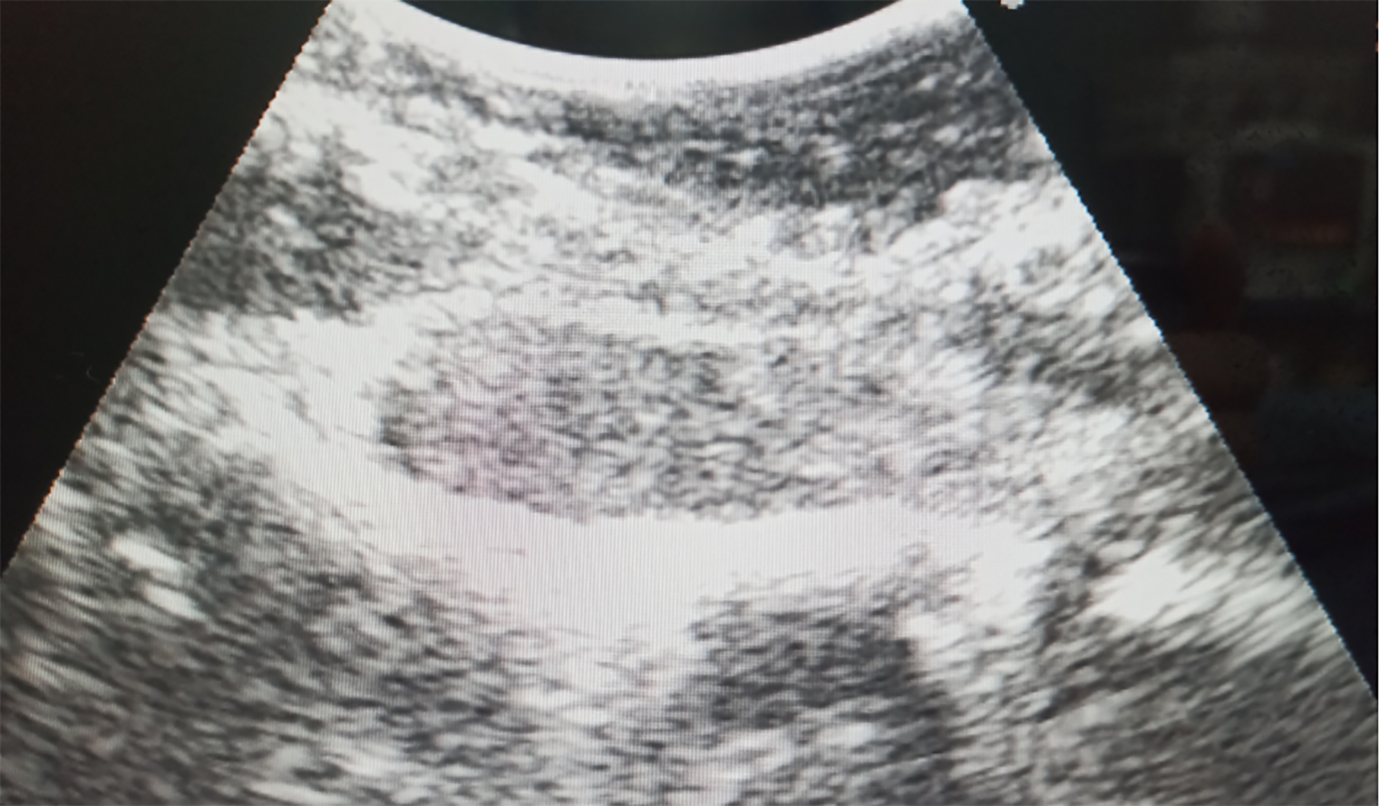
Figure 4 Intraoperative Ultrasonography for Localization of the Lesion Intraoperative ultrasonography precisely localized the lesion within the third vertebral body plane, situated along the ventral aspect of the spinal canal.
The lesion exhibited an elliptical shape and displayed a low signal echo, indicating its distinct characteristics.
- Citation: Zhou JG. Diagnosis and surgical challenges of extremely severe head and lumbar disc herniation in young patients: A case report. World J Clin Cases 2025; 13(16): 103373
- URL: https://www.wjgnet.com/2307-8960/full/v13/i16/103373.htm
- DOI: https://dx.doi.org/10.12998/wjcc.v13.i16.103373


