Copyright
©The Author(s) 2025.
World J Clin Cases. Jun 6, 2025; 13(16): 101665
Published online Jun 6, 2025. doi: 10.12998/wjcc.v13.i16.101665
Published online Jun 6, 2025. doi: 10.12998/wjcc.v13.i16.101665
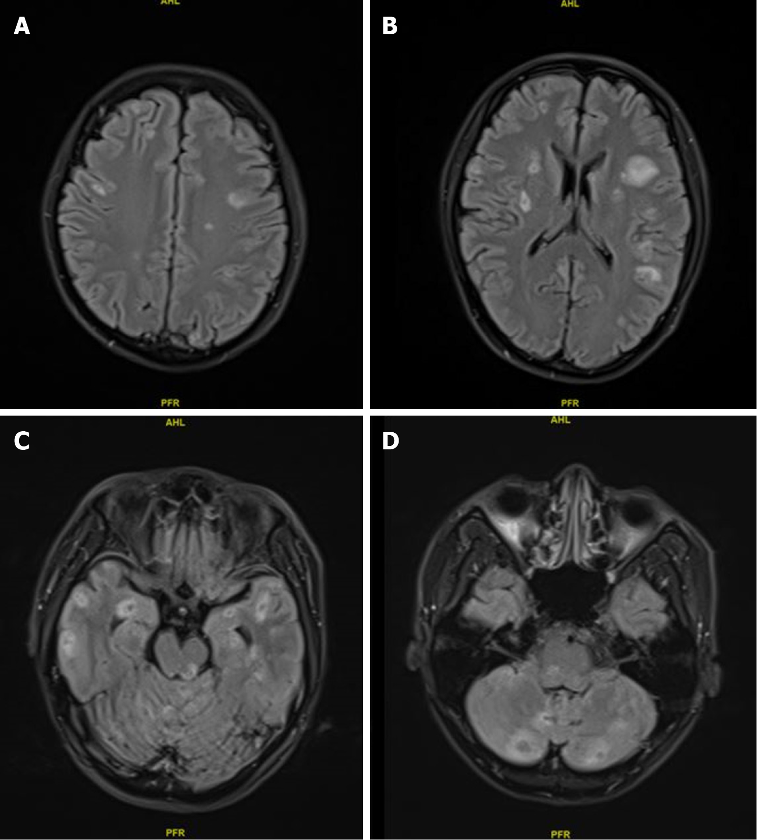
Figure 2 Magnetic resonance imaging of multiple intracranial metastases after first-line chemotherapy.
A: Magnetic resonance imaging (MRI) T1-weighted image revealed multiple lesions in the frontal and parietal regions; B: MRI T1-weighted image revealed multiple lesions in the basal ganglia; C: MRI T1-weighted image revealed multiple lesions in the temporal lobe and brainstem; D: MRI T1-weighted image revealed multiple lesions in the brainstem and cerebellum.
- Citation: Li F, Shen F. Metastatic pancreatic cancer with activating BRAF V600E mutations: A case report. World J Clin Cases 2025; 13(16): 101665
- URL: https://www.wjgnet.com/2307-8960/full/v13/i16/101665.htm
- DOI: https://dx.doi.org/10.12998/wjcc.v13.i16.101665


