Copyright
©The Author(s) 2025.
World J Clin Cases. Jun 6, 2025; 13(16): 101665
Published online Jun 6, 2025. doi: 10.12998/wjcc.v13.i16.101665
Published online Jun 6, 2025. doi: 10.12998/wjcc.v13.i16.101665
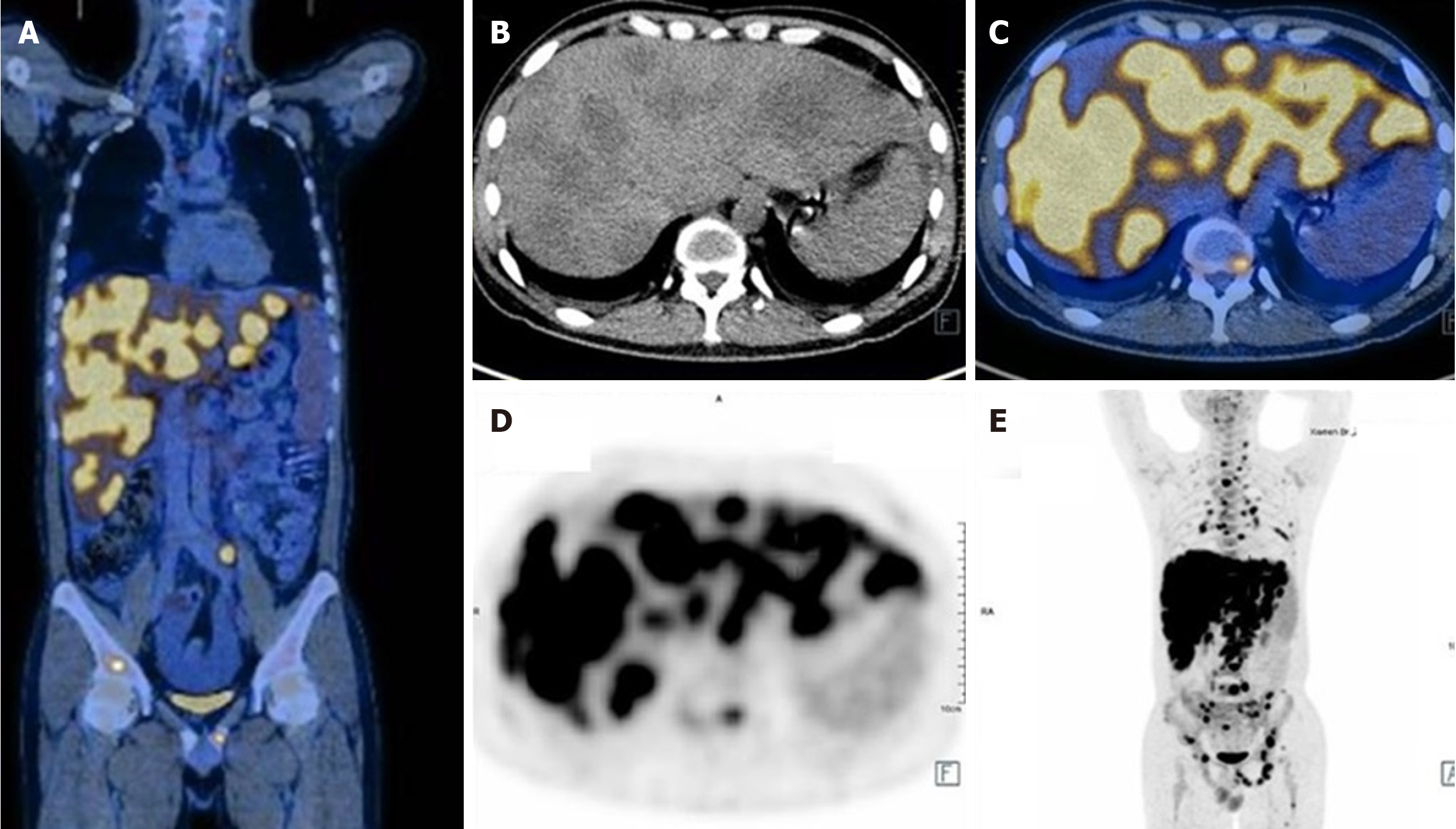
Figure 1 Positron emission tomography/computed tomography of whole body and abdomen before treatment.
A: Coronal bitmap of positron emission tomography/computed tomography (PET/CT) fusion image revealed high tumor burden with malignant lesions involving pancreas, liver, multiple bones, and multiple lymph nodes (bilateral neck, clavicle, mediastinum, bilateral hilum of lung, abdomen, pelvis and retroperitoneum; B: CT images revealed multiple low-density lesions in the liver; C: Cross-sectional PET/CT fusion image revealed multiple lesions with elevated 18F-fluorodeoxyglucose metabolism in liver and lumbar vertebra; D: PET image revealed multiple lesions with elevated 18F-fluorodeoxyglucose metabolism in liver and lumbar vertebra; E: Maximum intensity projection image revealed high tumor burden with malignant lesions involving pancreas, liver, multiple bones, and multiple lymph nodes (bilateral neck, clavicle, mediastinum, bilateral hilum of lung, abdomen, pelvis and retroperitoneum.
- Citation: Li F, Shen F. Metastatic pancreatic cancer with activating BRAF V600E mutations: A case report. World J Clin Cases 2025; 13(16): 101665
- URL: https://www.wjgnet.com/2307-8960/full/v13/i16/101665.htm
- DOI: https://dx.doi.org/10.12998/wjcc.v13.i16.101665


