Copyright
©The Author(s) 2025.
World J Clin Cases. May 26, 2025; 13(15): 101272
Published online May 26, 2025. doi: 10.12998/wjcc.v13.i15.101272
Published online May 26, 2025. doi: 10.12998/wjcc.v13.i15.101272
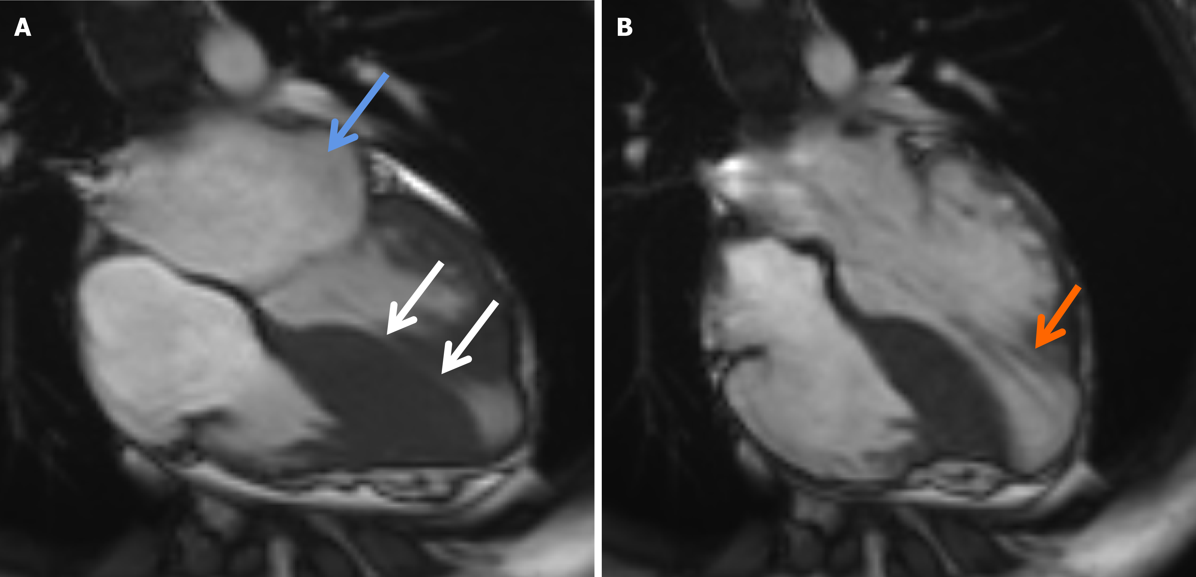
Figure 4 The cardiac magnetic resonance imaging findings of the proband.
A: The cardiac magnetic resonance imaging (CMR) found enlargement of left atrium (shown by the blue arrow), asymmetric hypertrophy of ventricular septum (shown by the white arrows), thinning of the bottom of ventricular septal base and left ventricle free wall; B: An apical ventricular aneurysm (about 24.6 mm × 29.8 mm) was shown by CMR (the orange arrow).
- Citation: Hong Y, Fan Z, Guo Y, Ma HH, Zeng SZ, Xi HT, Yang J, Luo K, Luo R, Li XP. MYH7 mutation in a pedigree with familial dilated hypertrophic cardiomyopathy: A case report and review of literature. World J Clin Cases 2025; 13(15): 101272
- URL: https://www.wjgnet.com/2307-8960/full/v13/i15/101272.htm
- DOI: https://dx.doi.org/10.12998/wjcc.v13.i15.101272


