Copyright
©The Author(s) 2025.
World J Clin Cases. Jan 6, 2025; 13(1): 99746
Published online Jan 6, 2025. doi: 10.12998/wjcc.v13.i1.99746
Published online Jan 6, 2025. doi: 10.12998/wjcc.v13.i1.99746
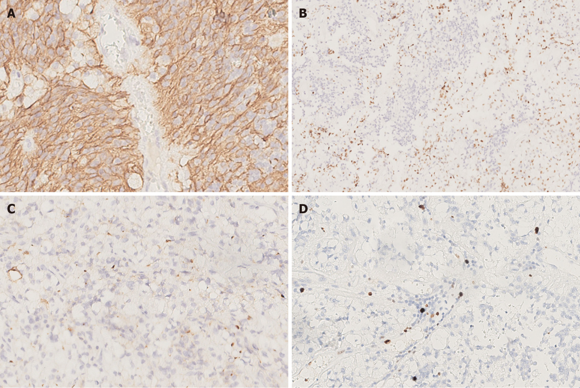
Figure 3 Immunohistochemistry findings.
A: The tumor cells were positive for GFAP [Immunohistochemistry (IHC) × 400]; B: Individual cells were positive for Olig-2 (IHC × 200); C: Significant punctate intracytoplasmic EMA immunoreactivity was observed (IHC × 400); D: The Ki-67 proliferation index was about 5% (IHC × 400).
- Citation: Zhao XY, Yu JH, Wang YH, Liu YX, Xu L, Fu L, Yi N. Lipomatous ependymoma with ZFTA: RELA fusion-positive: A case report. World J Clin Cases 2025; 13(1): 99746
- URL: https://www.wjgnet.com/2307-8960/full/v13/i1/99746.htm
- DOI: https://dx.doi.org/10.12998/wjcc.v13.i1.99746


