Copyright
©The Author(s) 2025.
World J Clin Cases. Jan 6, 2025; 13(1): 99746
Published online Jan 6, 2025. doi: 10.12998/wjcc.v13.i1.99746
Published online Jan 6, 2025. doi: 10.12998/wjcc.v13.i1.99746
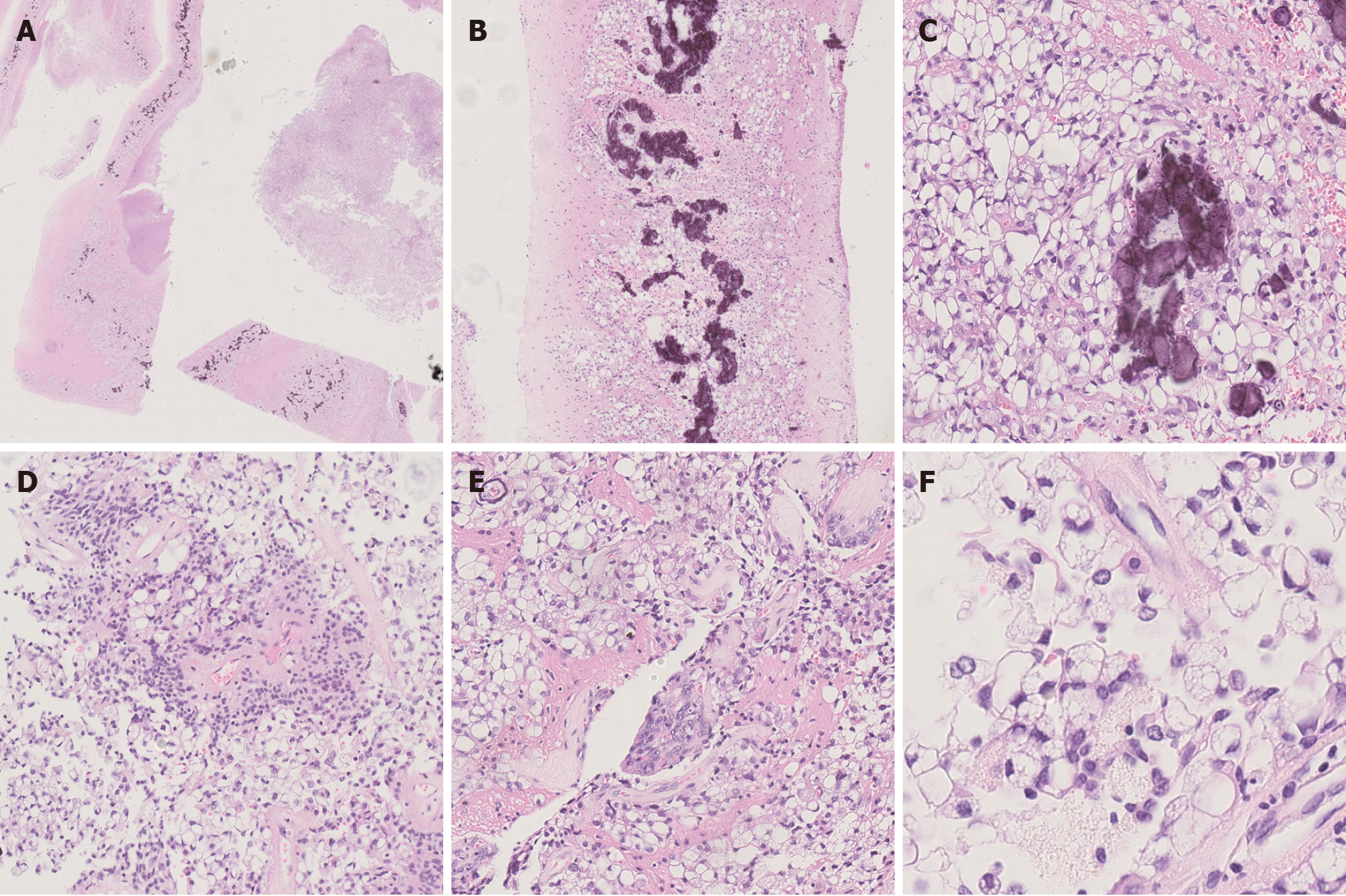
Figure 2 Histopathological findings.
A: The tissue boundary in the cystic wall-like structure was clear [hematoxylin and eosin (HE) × 40]; B: Calcifications were seen on the cyst wall (HE × 100); C: Large vacuoles were seen in the cytoplasm of tumor cells on the cyst wall (HE × 400); D: Characteristic perivascular pseudorosettes (HE × 200); E: Vascular endothelial cell proliferation was seen, resembling a glomerular structure (HE × 200); F: Vacuoles in the cytoplasm pushed the crescentic nucleus, similar to signet ring cells (HE × 1000).
- Citation: Zhao XY, Yu JH, Wang YH, Liu YX, Xu L, Fu L, Yi N. Lipomatous ependymoma with ZFTA: RELA fusion-positive: A case report. World J Clin Cases 2025; 13(1): 99746
- URL: https://www.wjgnet.com/2307-8960/full/v13/i1/99746.htm
- DOI: https://dx.doi.org/10.12998/wjcc.v13.i1.99746


