Copyright
©The Author(s) 2025.
World J Clin Cases. Jan 6, 2025; 13(1): 99746
Published online Jan 6, 2025. doi: 10.12998/wjcc.v13.i1.99746
Published online Jan 6, 2025. doi: 10.12998/wjcc.v13.i1.99746
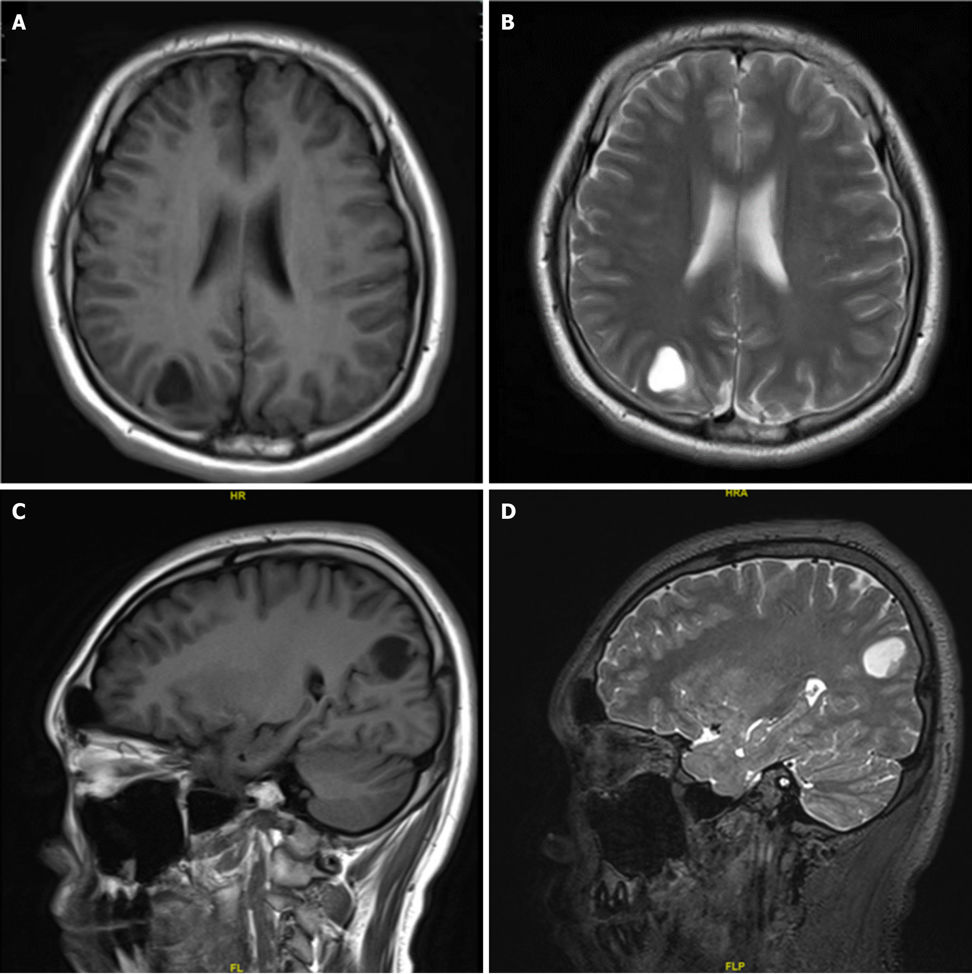
Figure 1 Magnetic resonance imaging showed a cystic mass of about 1.
9 cm × 1.5 cm × 1.9 cm under the cortex of the right parietal occipital lobe, with a uniform long T1 and long T2 signal. The cystic mass was regular, the edges were clear and smooth, and the surrounding brain tissue was slightly compressed. The remaining brain parenchyma showed no abnormal signal shadows. A: Axial T1; B: Axial T2; C: Sagittal T1; D: Sagittal T2.
- Citation: Zhao XY, Yu JH, Wang YH, Liu YX, Xu L, Fu L, Yi N. Lipomatous ependymoma with ZFTA: RELA fusion-positive: A case report. World J Clin Cases 2025; 13(1): 99746
- URL: https://www.wjgnet.com/2307-8960/full/v13/i1/99746.htm
- DOI: https://dx.doi.org/10.12998/wjcc.v13.i1.99746


