Copyright
©The Author(s) 2024.
World J Clin Cases. Mar 26, 2024; 12(9): 1677-1684
Published online Mar 26, 2024. doi: 10.12998/wjcc.v12.i9.1677
Published online Mar 26, 2024. doi: 10.12998/wjcc.v12.i9.1677
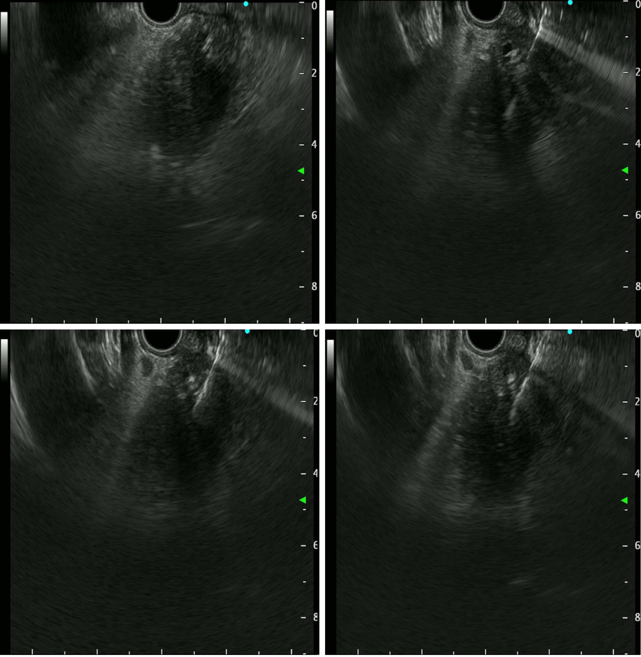
Figure 3 Endoscopic ultrasound-guided tissue sampling.
Endoscopic ultrasound (EUS) showed a 36.0 mm × 32.9 mm sized ill-defined relative heterogeneous hypoechoic mass in the pancreatic head. EUS-guided tissue sampling (EUS-TS) was performed using a 22-gauge EZ Shot (EZ Shot 3 Plus; Olympus, Tokyo, Japan) using the fanning method.
- Citation: Kim KH, Park CH, Cho E, Lee Y. Endoscopic ultrasound-guided tissue sampling induced pancreatic duct leak resolved by the placement of a pancreatic stent: A case report. World J Clin Cases 2024; 12(9): 1677-1684
- URL: https://www.wjgnet.com/2307-8960/full/v12/i9/1677.htm
- DOI: https://dx.doi.org/10.12998/wjcc.v12.i9.1677


