Copyright
©The Author(s) 2024.
World J Clin Cases. Mar 16, 2024; 12(8): 1497-1503
Published online Mar 16, 2024. doi: 10.12998/wjcc.v12.i8.1497
Published online Mar 16, 2024. doi: 10.12998/wjcc.v12.i8.1497
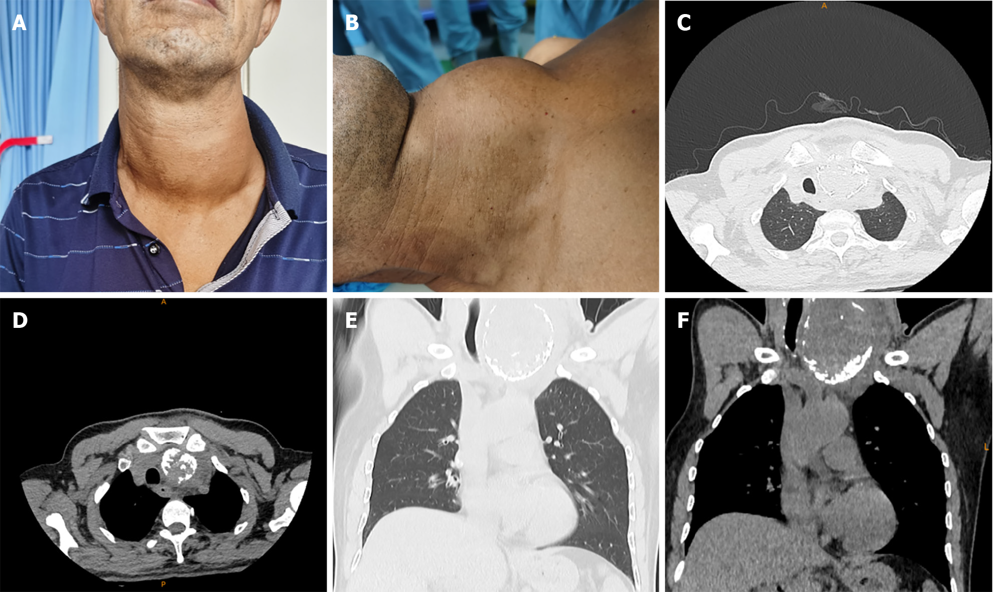
Figure 1 Neck signs and computed tomography scan imaging.
A: Front view of the neck mass; B: Lateral view of the neck mass; C and D: Computed tomography (CT) lung window and mediastinal window: Transverse images show a huge thyroid nodule and deviation of compressed trachea and esophagus to right posterior side; E and F: CT lung window and mediastinal window: Coronal images show a huge thyroid mass and deviation of compressed trachea to right side.
- Citation: Liu JB, Zhang SL, Jiang WL, Sun HK, Yang HC. Chronic infectious unilateral giant thyroid cyst related to diabetes mellitus: A case report. World J Clin Cases 2024; 12(8): 1497-1503
- URL: https://www.wjgnet.com/2307-8960/full/v12/i8/1497.htm
- DOI: https://dx.doi.org/10.12998/wjcc.v12.i8.1497


