Copyright
©The Author(s) 2024.
World J Clin Cases. Mar 16, 2024; 12(8): 1487-1496
Published online Mar 16, 2024. doi: 10.12998/wjcc.v12.i8.1487
Published online Mar 16, 2024. doi: 10.12998/wjcc.v12.i8.1487
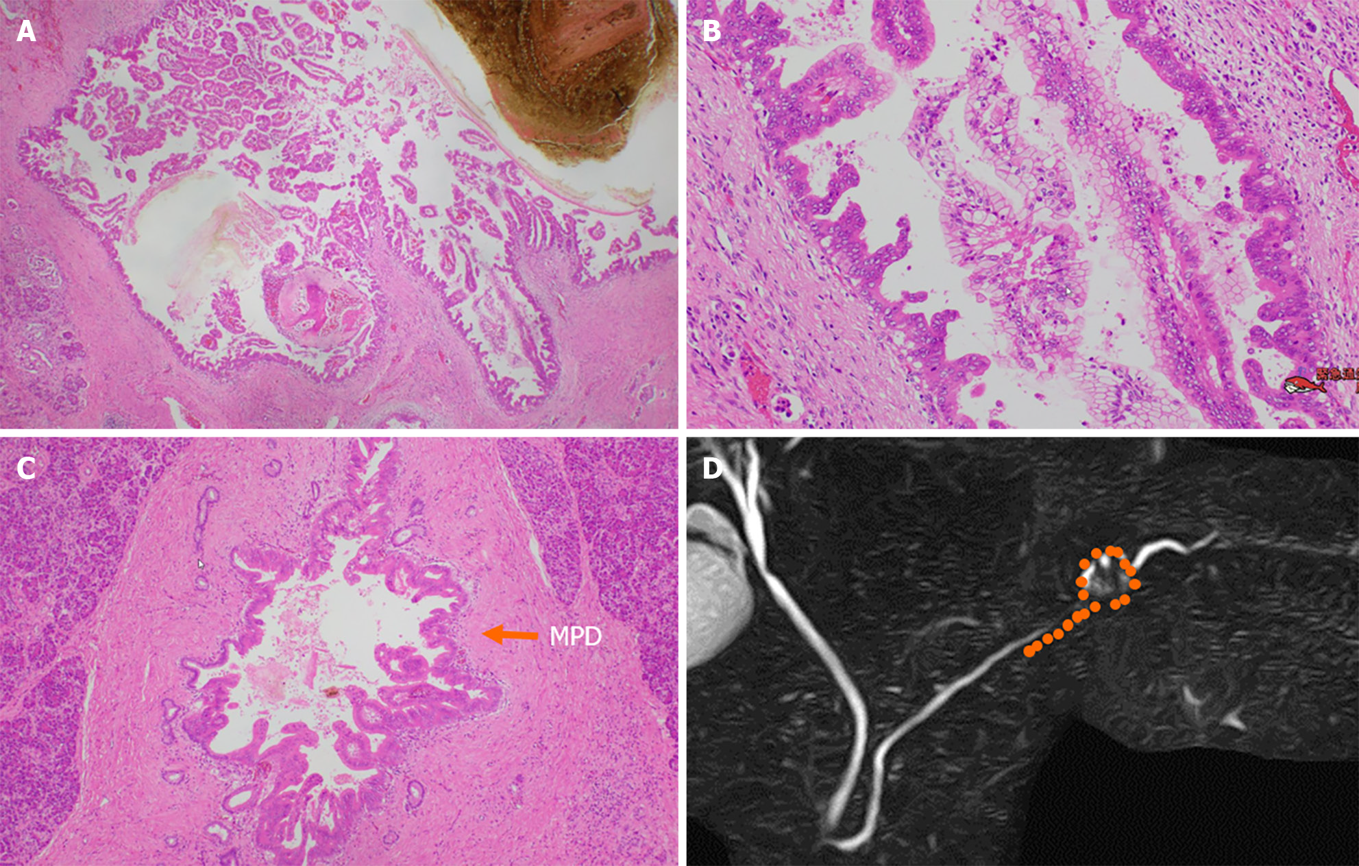
Figure 8 Pathological findings.
A and B: Low-papillary and pseudopapillary adenocarcinoma cells were spread in the cyst wall; C: Malignant cells spread to the main pancreatic duct (arrow) and branch duct, in a 20-mm range toward the pancreatic body; D: Distribution of high-grade pancreatic intraepithelial neoplasia (orange points). MPD: Main pancreatic duct.
- Citation: Furuya N, Yamaguchi A, Kato N, Sugata S, Hamada T, Mizumoto T, Tamaru Y, Kusunoki R, Kuwai T, Kouno H, Kuraoka K, Shibata Y, Tazuma S, Sudo T, Kohno H, Oka S. High-grade pancreatic intraepithelial neoplasia diagnosed based on changes in magnetic resonance cholangiopancreatography findings: A case report. World J Clin Cases 2024; 12(8): 1487-1496
- URL: https://www.wjgnet.com/2307-8960/full/v12/i8/1487.htm
- DOI: https://dx.doi.org/10.12998/wjcc.v12.i8.1487


