Copyright
©The Author(s) 2024.
World J Clin Cases. Mar 16, 2024; 12(8): 1487-1496
Published online Mar 16, 2024. doi: 10.12998/wjcc.v12.i8.1487
Published online Mar 16, 2024. doi: 10.12998/wjcc.v12.i8.1487
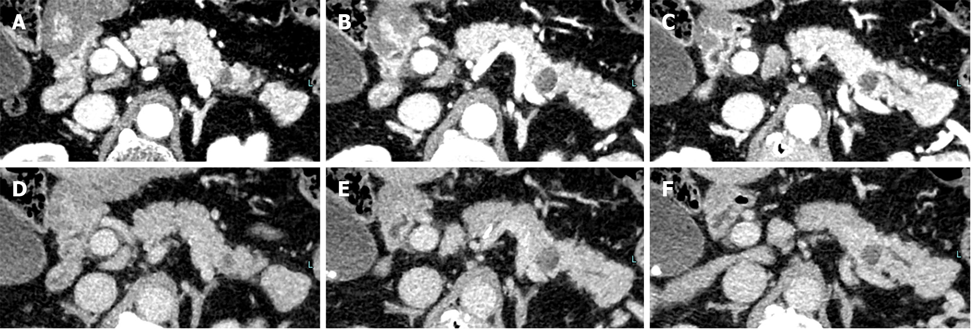
Figure 5 Computed tomography at 24 months revealed no solid space-occupying lesion but the pancreatic tail cyst has grown to 10 mm and the caudal main pancreatic duct was slightly dilated.
A-C: Pancreatic parenchymal phase; D-F: Equilibrium phase.
- Citation: Furuya N, Yamaguchi A, Kato N, Sugata S, Hamada T, Mizumoto T, Tamaru Y, Kusunoki R, Kuwai T, Kouno H, Kuraoka K, Shibata Y, Tazuma S, Sudo T, Kohno H, Oka S. High-grade pancreatic intraepithelial neoplasia diagnosed based on changes in magnetic resonance cholangiopancreatography findings: A case report. World J Clin Cases 2024; 12(8): 1487-1496
- URL: https://www.wjgnet.com/2307-8960/full/v12/i8/1487.htm
- DOI: https://dx.doi.org/10.12998/wjcc.v12.i8.1487


