Copyright
©The Author(s) 2024.
World J Clin Cases. Mar 16, 2024; 12(8): 1487-1496
Published online Mar 16, 2024. doi: 10.12998/wjcc.v12.i8.1487
Published online Mar 16, 2024. doi: 10.12998/wjcc.v12.i8.1487
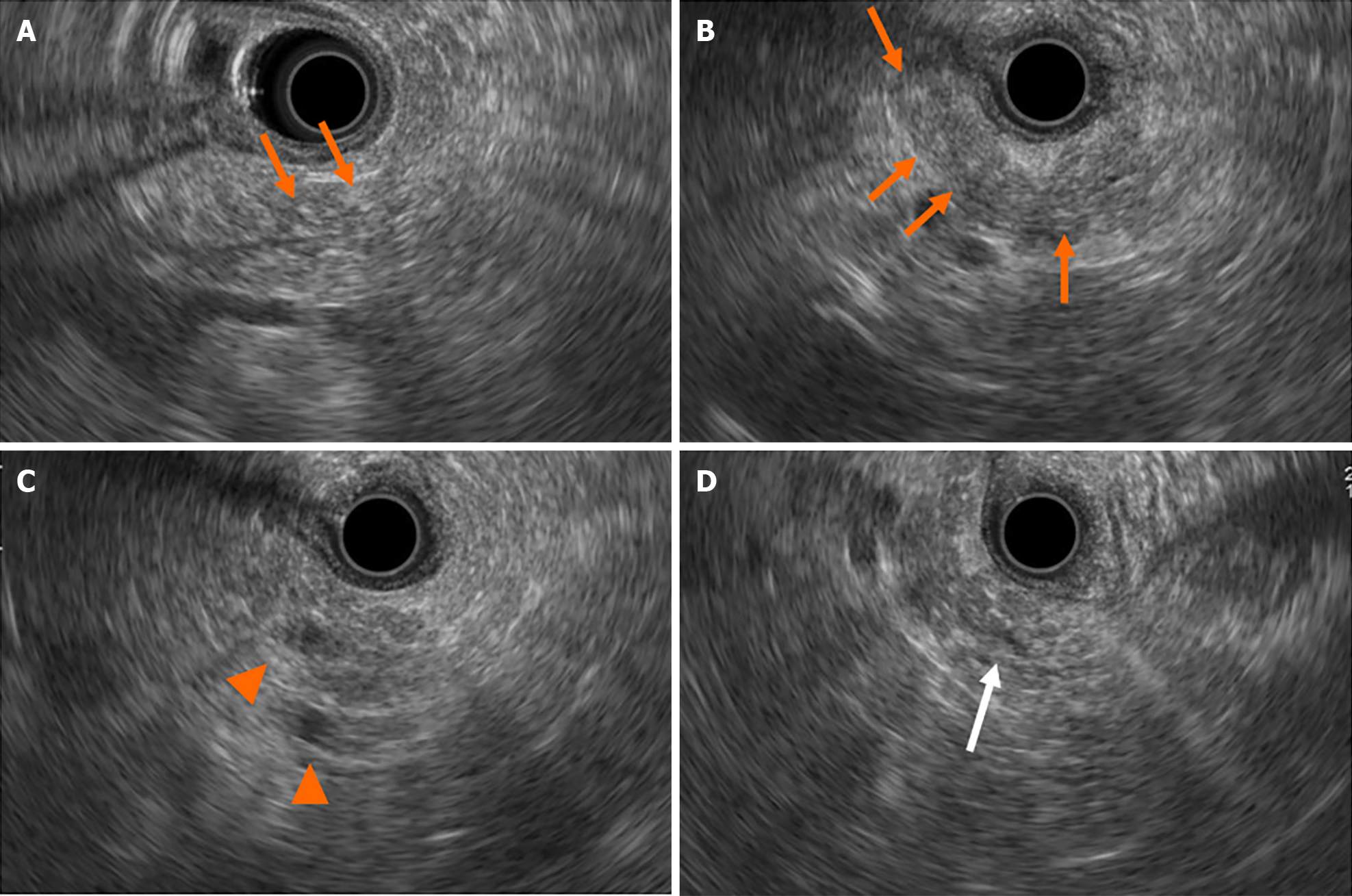
Figure 2 Findings of endoscopic ultrasonography at first visit to our institute.
A and B: There were diffuse high echoic spots (orange arrows) in the whole pancreas, indicating early chronic pancreatitis; C: Small round cysts (arrowheads) were found in the pancreatic tail; D: There was no caudal main pancreatic duct dilatation in the pancreatic tail (white arrow).
- Citation: Furuya N, Yamaguchi A, Kato N, Sugata S, Hamada T, Mizumoto T, Tamaru Y, Kusunoki R, Kuwai T, Kouno H, Kuraoka K, Shibata Y, Tazuma S, Sudo T, Kohno H, Oka S. High-grade pancreatic intraepithelial neoplasia diagnosed based on changes in magnetic resonance cholangiopancreatography findings: A case report. World J Clin Cases 2024; 12(8): 1487-1496
- URL: https://www.wjgnet.com/2307-8960/full/v12/i8/1487.htm
- DOI: https://dx.doi.org/10.12998/wjcc.v12.i8.1487


