Copyright
©The Author(s) 2024.
World J Clin Cases. Mar 16, 2024; 12(8): 1467-1473
Published online Mar 16, 2024. doi: 10.12998/wjcc.v12.i8.1467
Published online Mar 16, 2024. doi: 10.12998/wjcc.v12.i8.1467
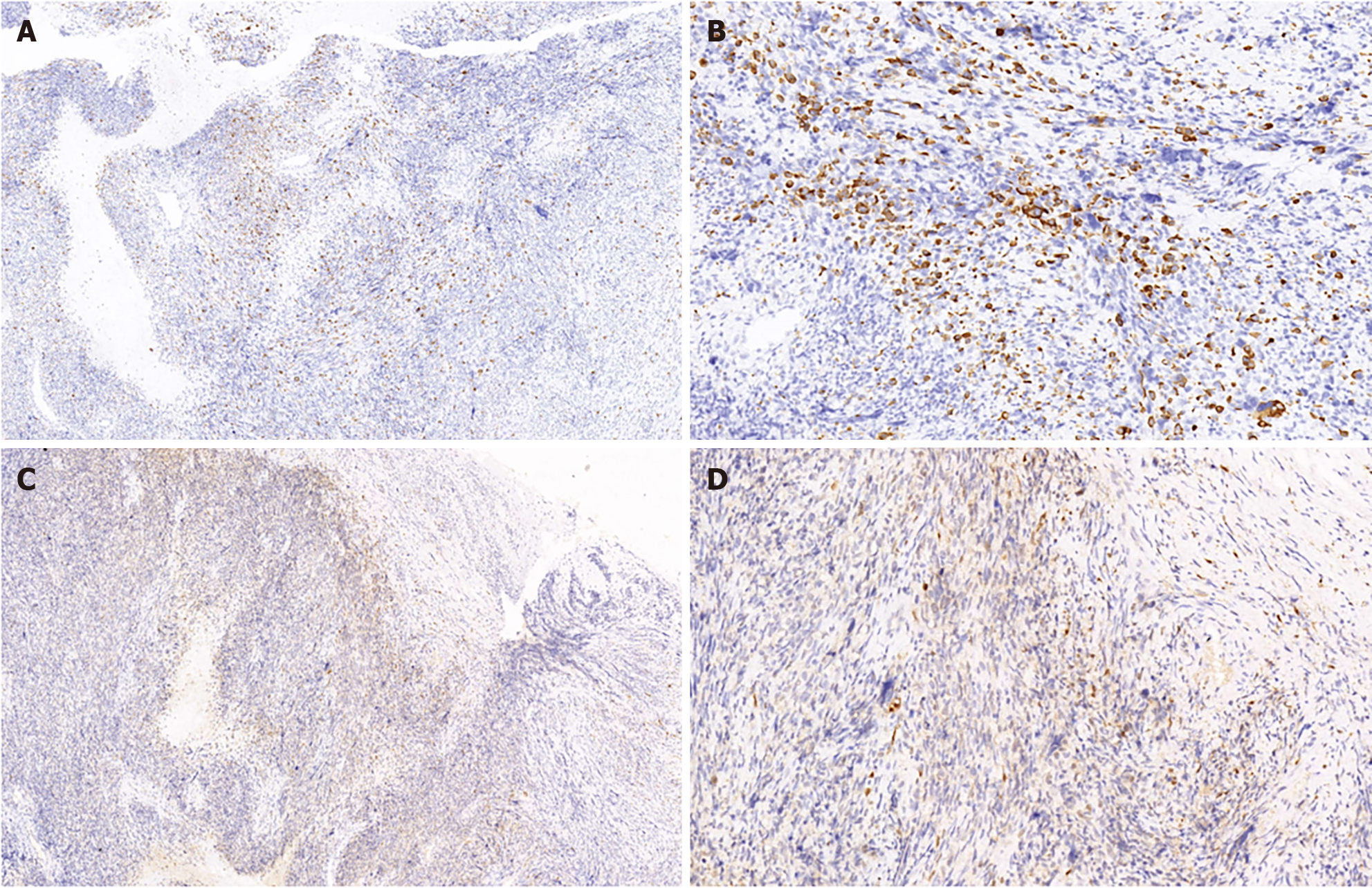
Figure 3 Immunohistochemical examination of the resected specimen.
A: Rhabdomyoblasts exhibited focal positivity for desmin (immunohistochemical staining, × 100); B: Desmin (immunohistochemical staining, × 300); C: S-100 was also detected in the malignant cells (immunohistochemical staining, × 100); D: S-100 (immunohistochemical staining, × 300).
- Citation: Yang HJ, Kim D, Lee WS, Oh SH. Malignant triton tumor in the abdominal wall: A case report. World J Clin Cases 2024; 12(8): 1467-1473
- URL: https://www.wjgnet.com/2307-8960/full/v12/i8/1467.htm
- DOI: https://dx.doi.org/10.12998/wjcc.v12.i8.1467


