Copyright
©The Author(s) 2024.
World J Clin Cases. Mar 16, 2024; 12(8): 1448-1453
Published online Mar 16, 2024. doi: 10.12998/wjcc.v12.i8.1448
Published online Mar 16, 2024. doi: 10.12998/wjcc.v12.i8.1448
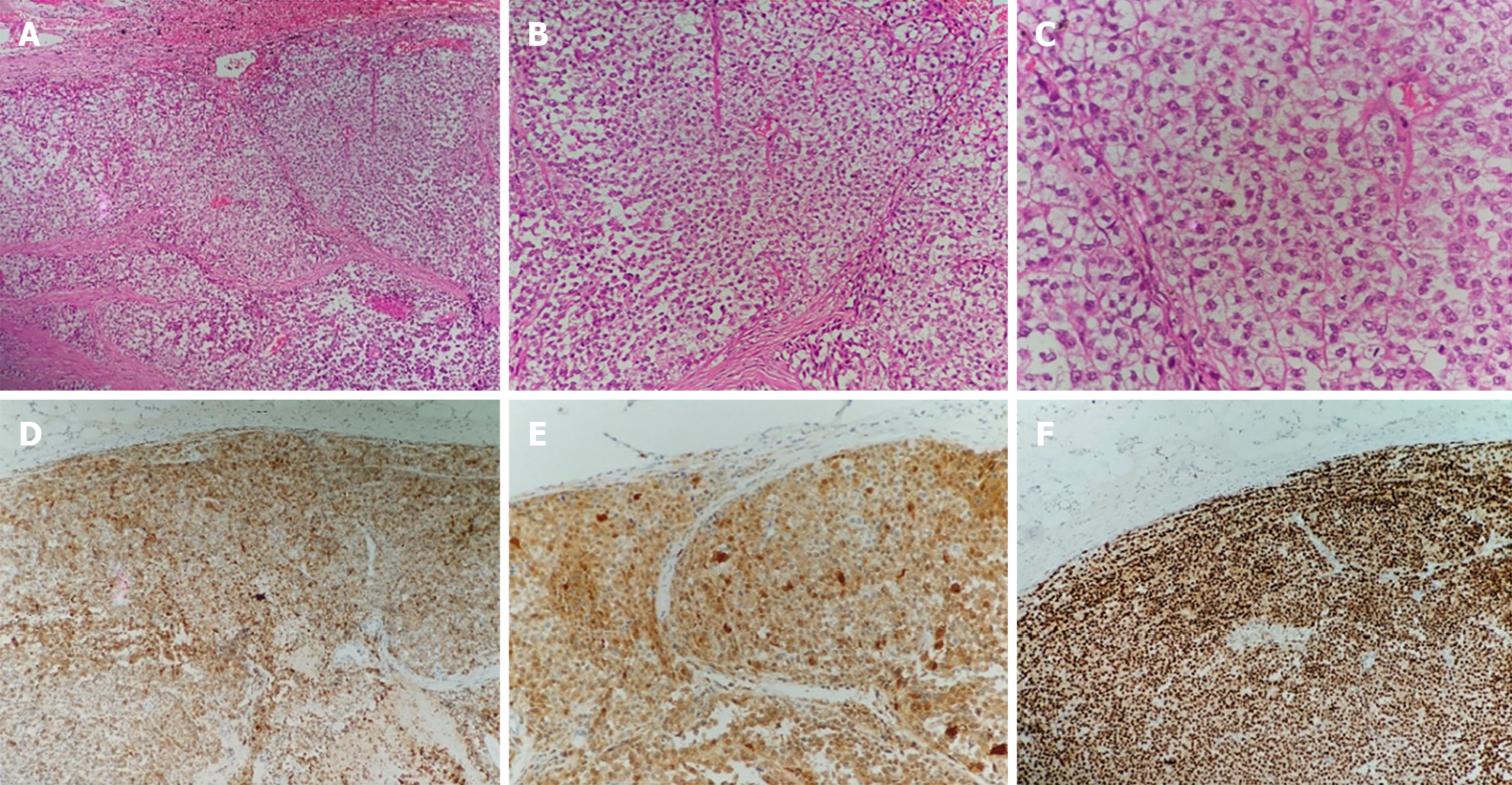
Figure 3 The malignant tumor of the pancreatic body was accompanied by massive necrosis.
A and B: Hematoxylin and eosin (H&E) showed tumor cells arranged in nests (A; × 10), with fibrous and vascular separation around the nests (B; × 20); C: The cells were oval or polygonal, with obvious nucleoli, and some cytoplasm was lightly stained or vacuolated (H&E, × 40); D-F: No metastasis was found in the surrounding lymph nodes and no tumor involvement was found in the resection margin. Immunohistochemical results: AE1/3 (-), CD56 (-), CgA (-), CK8/18(-), HMB45(+), Melan-A (+) (D), Ki-67 (60%+), S-100 (+)(E), SOX10 (+)(F), syn (-), β-catenin (+), PR (-).
- Citation: Liu YJ, Zou C, Wu YY. Metastatic clear cell sarcoma of the pancreas: A rare case report. World J Clin Cases 2024; 12(8): 1448-1453
- URL: https://www.wjgnet.com/2307-8960/full/v12/i8/1448.htm
- DOI: https://dx.doi.org/10.12998/wjcc.v12.i8.1448


