Copyright
©The Author(s) 2024.
World J Clin Cases. Mar 16, 2024; 12(8): 1448-1453
Published online Mar 16, 2024. doi: 10.12998/wjcc.v12.i8.1448
Published online Mar 16, 2024. doi: 10.12998/wjcc.v12.i8.1448
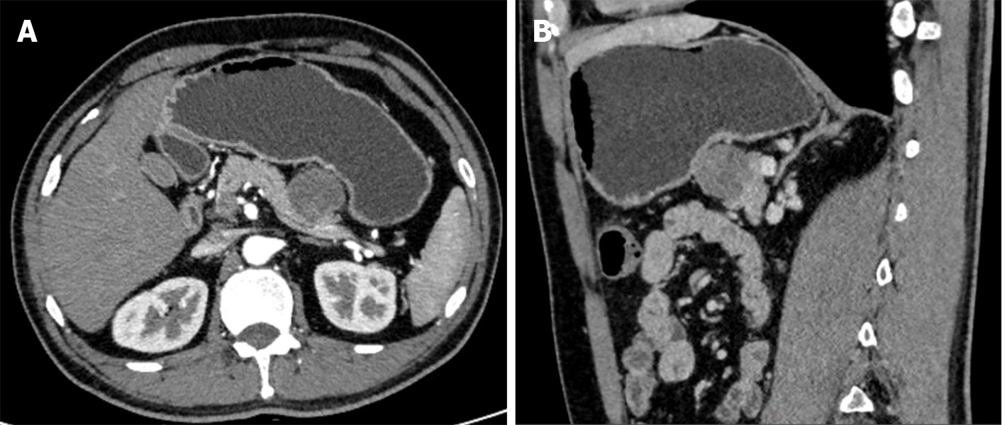
Figure 1 Abdominal computer tomograph showed a 3.
2 cm × 3.0 cm round lesion in the tail of the pancreas, which was uneven and mildly enhanced under enhanced scan. A: The boundary between the posterior margin and the pancreas was not clear, and was not combined with the pancreatic duct; B: Multiple lymph nodes were also seen in the mesentery area, with the width of about 1.3 cm for the largest one.
- Citation: Liu YJ, Zou C, Wu YY. Metastatic clear cell sarcoma of the pancreas: A rare case report. World J Clin Cases 2024; 12(8): 1448-1453
- URL: https://www.wjgnet.com/2307-8960/full/v12/i8/1448.htm
- DOI: https://dx.doi.org/10.12998/wjcc.v12.i8.1448


