Copyright
©The Author(s) 2024.
World J Clin Cases. Mar 16, 2024; 12(8): 1422-1429
Published online Mar 16, 2024. doi: 10.12998/wjcc.v12.i8.1422
Published online Mar 16, 2024. doi: 10.12998/wjcc.v12.i8.1422
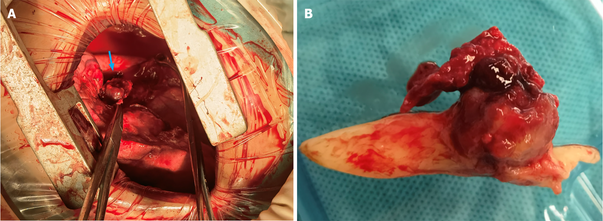
Figure 2 Intraoperative images.
A: An anterolateral incision facilitated sleeve resection of the left lower lobe and systematic mediastinal lymph node dissection. The image depicts the hyperinflated left upper lung, the severed left lower lobe bronchus, and a 2 cm dark red spherical tumor obstructing the bronchial opening in the left lower lobe while infiltrating the bronchial wall (indicated by a blue arrow); B: The tumor was accompanied by a white, tongue-shaped, clotted sputum plug extending into the left upper bronchus and obstructing a substantial portion of the lumen.
- Citation: Yu XH, Wang WX, Yang DS, Gong LH. Left lower lobe sleeve resection for the clear cell variant of pulmonary mucoepidermoid carcinoma: A case report. World J Clin Cases 2024; 12(8): 1422-1429
- URL: https://www.wjgnet.com/2307-8960/full/v12/i8/1422.htm
- DOI: https://dx.doi.org/10.12998/wjcc.v12.i8.1422


