Copyright
©The Author(s) 2024.
World J Clin Cases. Mar 16, 2024; 12(8): 1422-1429
Published online Mar 16, 2024. doi: 10.12998/wjcc.v12.i8.1422
Published online Mar 16, 2024. doi: 10.12998/wjcc.v12.i8.1422
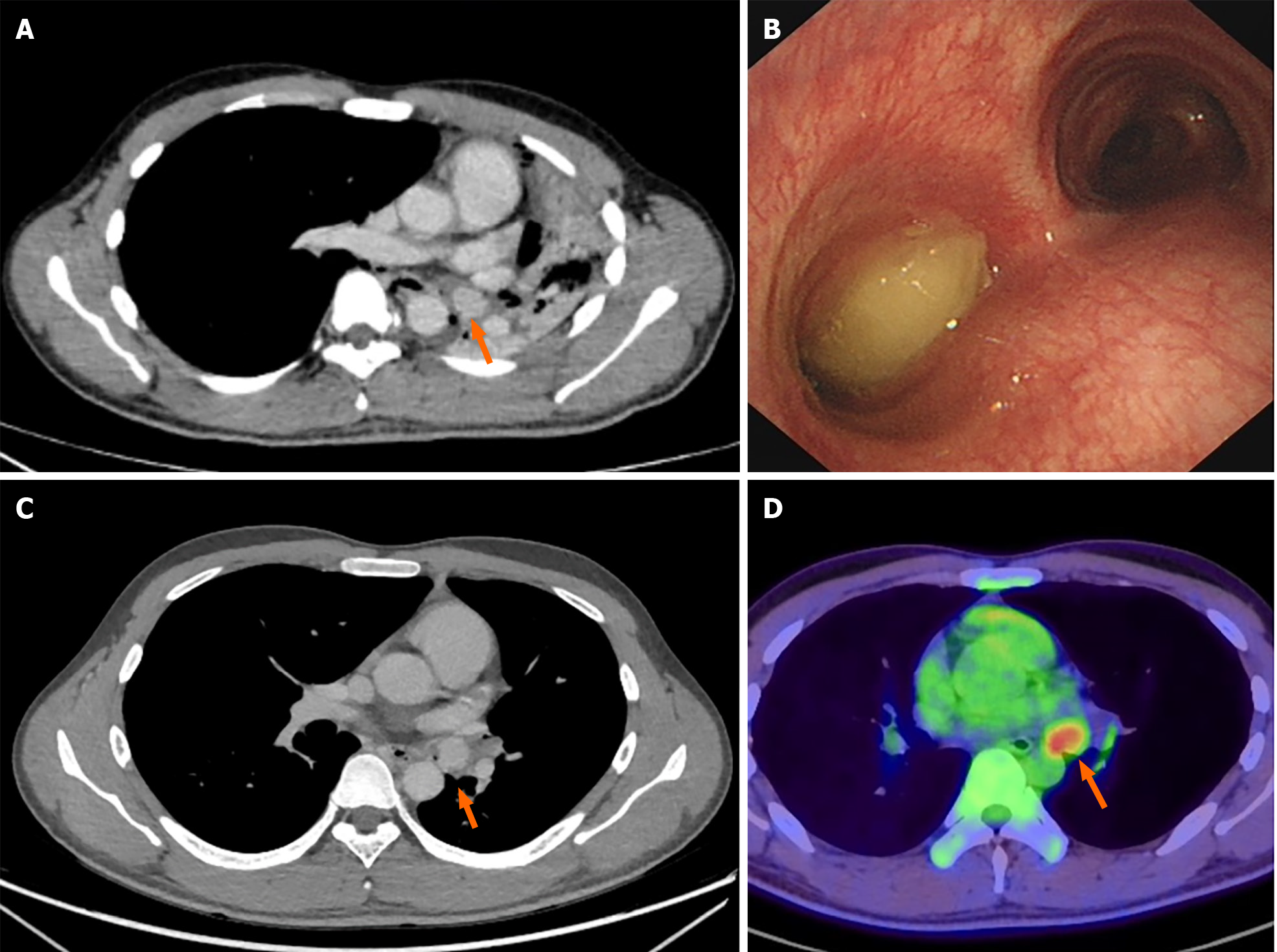
Figure 1 Preoperative patient examination.
A: Chest computed tomography (CT) scan with enhanced imaging revealed a 2-cm soft tissue density nodule situated in the left main bronchus, resulting in complete atelectasis of the left lung; B: Electronic bronchoscopy image showed a polypoid tumor obstructing the left main bronchus, featuring a surface covered with gray yellow necrotic material; C: CT imaging demonstrated signs of recruitment in the left upper lung after endoscopic treatment, while the mass in the left main bronchus persisted; D: Whole-body positron emission tomography/CT scan revealed metabolically active lesions indicative of malignancy, with the tumor location highlighted by a red arrow.
- Citation: Yu XH, Wang WX, Yang DS, Gong LH. Left lower lobe sleeve resection for the clear cell variant of pulmonary mucoepidermoid carcinoma: A case report. World J Clin Cases 2024; 12(8): 1422-1429
- URL: https://www.wjgnet.com/2307-8960/full/v12/i8/1422.htm
- DOI: https://dx.doi.org/10.12998/wjcc.v12.i8.1422


