Copyright
©The Author(s) 2024.
World J Clin Cases. Mar 6, 2024; 12(7): 1326-1332
Published online Mar 6, 2024. doi: 10.12998/wjcc.v12.i7.1326
Published online Mar 6, 2024. doi: 10.12998/wjcc.v12.i7.1326
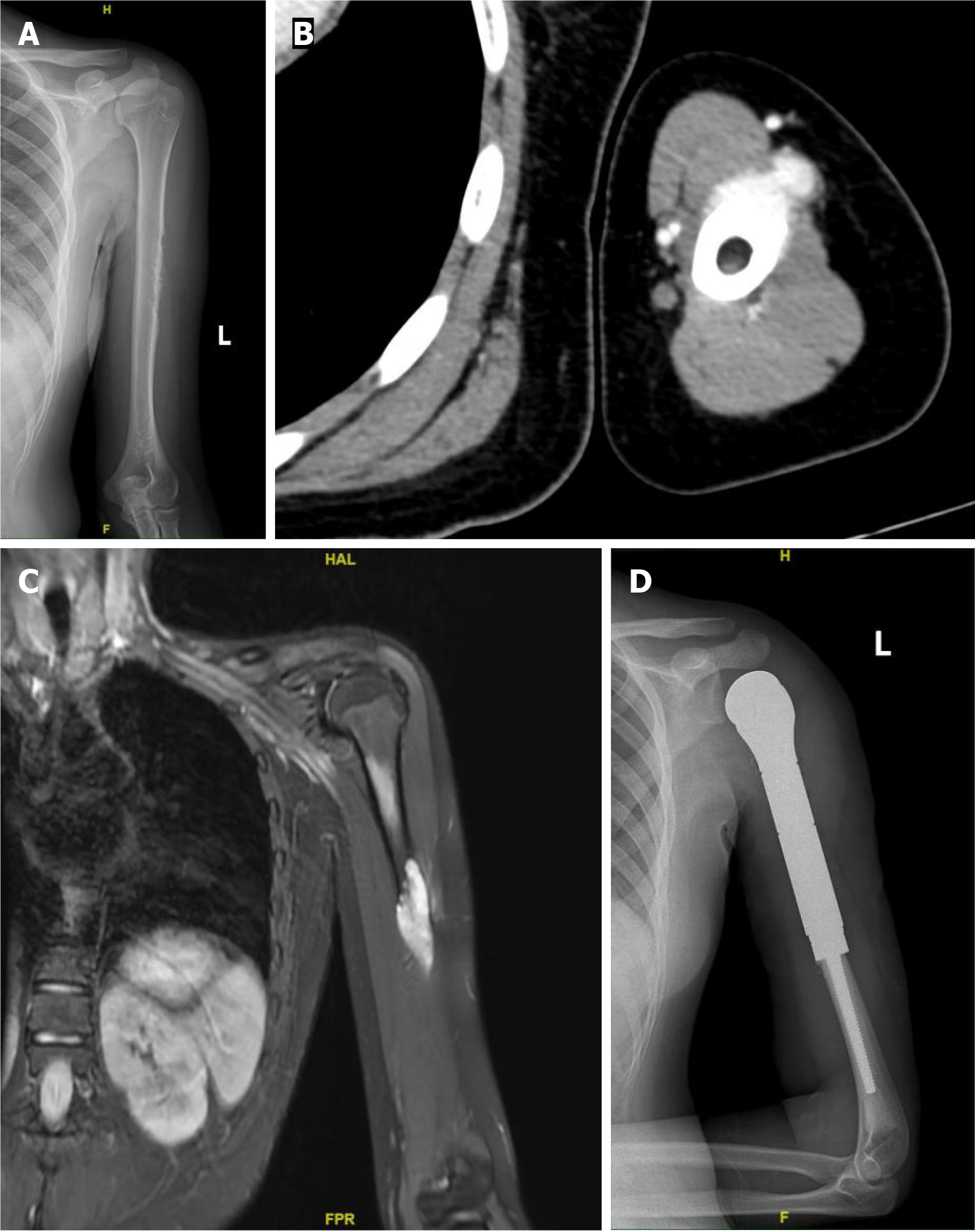
Figure 1 Imaging findings of the lesion.
A: X-ray reveals bone destruction and thinning of the corresponding segment of the cortical bone in the midshaft of the left femur; B: Computed tomography scan reveals roughness of the cortical bone in the middle segment of the left humerus, with surrounding soft tissue masses exhibiting a nodular morphology; C: Magnetic resonance imaging reveals a soft tissue mass surrounding the left humerus, which appears as a high signal on diffusion-weighted imaging; D: The postoperative X-ray indicates the presence of metallic internal fixation material in the surgical area of the left humerus.
- Citation: Zhou Y, Sun YW, Liu XY, Shen DH. Misdiagnosis of synovial sarcoma - cellular myofibroma with SRF-RELA gene fusion: A case report. World J Clin Cases 2024; 12(7): 1326-1332
- URL: https://www.wjgnet.com/2307-8960/full/v12/i7/1326.htm
- DOI: https://dx.doi.org/10.12998/wjcc.v12.i7.1326


