Copyright
©The Author(s) 2024.
World J Clin Cases. Mar 6, 2024; 12(7): 1284-1289
Published online Mar 6, 2024. doi: 10.12998/wjcc.v12.i7.1284
Published online Mar 6, 2024. doi: 10.12998/wjcc.v12.i7.1284
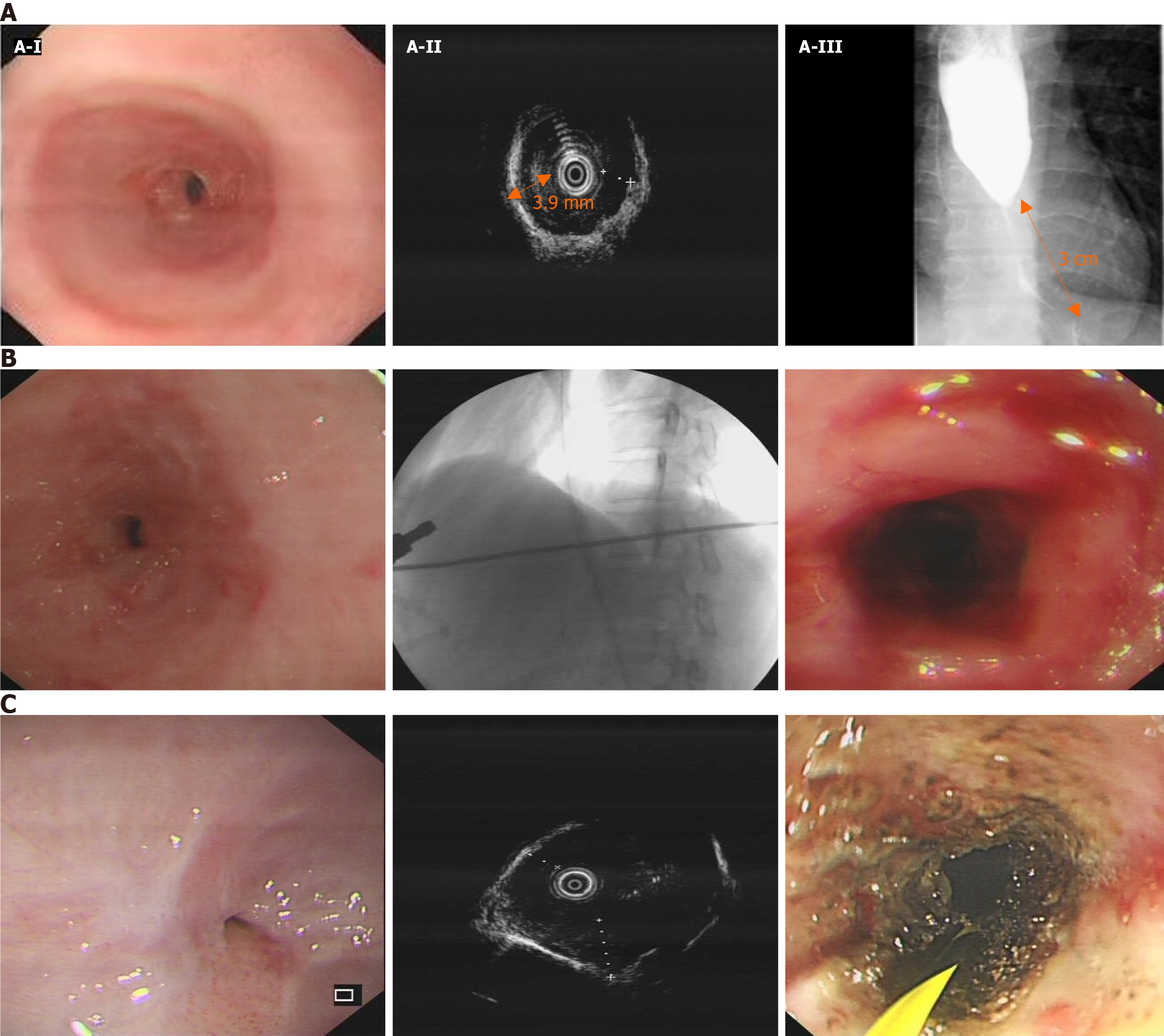
Figure 2 Treatment of esophageal stricture.
A: Assessment of esophageal stricture. The esophagus-gastro-duodenoscopy shows severe esophageal stricture with endoscopy unable to pass through (A-I); Endoscopic ultrasonography shows prominent thickening of the esophageal muscular layer, with the thickest part measuring approximately 3.9 mm (A-II); Esophagography shows severe lower esophageal stricture with a luminal diameter of 1.8 mm, involving a 3 cm length of the esophagus (A-III); B: A representative wire-guided endoscopic bougie dilations. The bougie is used to expand along with the wire; C: A representative ultrasonography-guided and wire-guided endoscopic incisional therapy. The needle knife is used to cut through the muscle layer along with the wire.
- Citation: Chen QN, Bai BQ, Xu Y, Mei Q, Liu XC. Sporadic gastrinoma with refractory benign esophageal stricture: A case report. World J Clin Cases 2024; 12(7): 1284-1289
- URL: https://www.wjgnet.com/2307-8960/full/v12/i7/1284.htm
- DOI: https://dx.doi.org/10.12998/wjcc.v12.i7.1284


