Copyright
©The Author(s) 2024.
World J Clin Cases. Mar 6, 2024; 12(7): 1284-1289
Published online Mar 6, 2024. doi: 10.12998/wjcc.v12.i7.1284
Published online Mar 6, 2024. doi: 10.12998/wjcc.v12.i7.1284
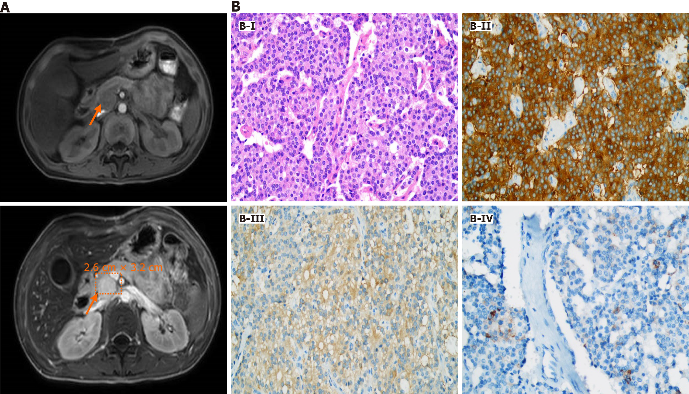
Figure 1 Diagnosis of gastrinoma.
A: Plain and enhanced magnetic resonance imaging scans of the pancreas showed a mass approximately 2.6 cm × 3.2 cm in size located at the head of the pancreas (indicated by orange arrows); B: Hematoxylin-eosin staining (× 400) and immunohistochemical stains (× 400) of the mass: The tumor cells, arranged into solid nests, exhibited abundant cytoplasm, fine and granulated chromatin, and inconspicuous nucleoli (B-I); Tumor cells were positive for synaptophysin (B-II), neuron-specific enolase (B-III), and gastrin (B-IV).
- Citation: Chen QN, Bai BQ, Xu Y, Mei Q, Liu XC. Sporadic gastrinoma with refractory benign esophageal stricture: A case report. World J Clin Cases 2024; 12(7): 1284-1289
- URL: https://www.wjgnet.com/2307-8960/full/v12/i7/1284.htm
- DOI: https://dx.doi.org/10.12998/wjcc.v12.i7.1284


