Copyright
©The Author(s) 2024.
World J Clin Cases. Feb 26, 2024; 12(6): 1157-1162
Published online Feb 26, 2024. doi: 10.12998/wjcc.v12.i6.1157
Published online Feb 26, 2024. doi: 10.12998/wjcc.v12.i6.1157
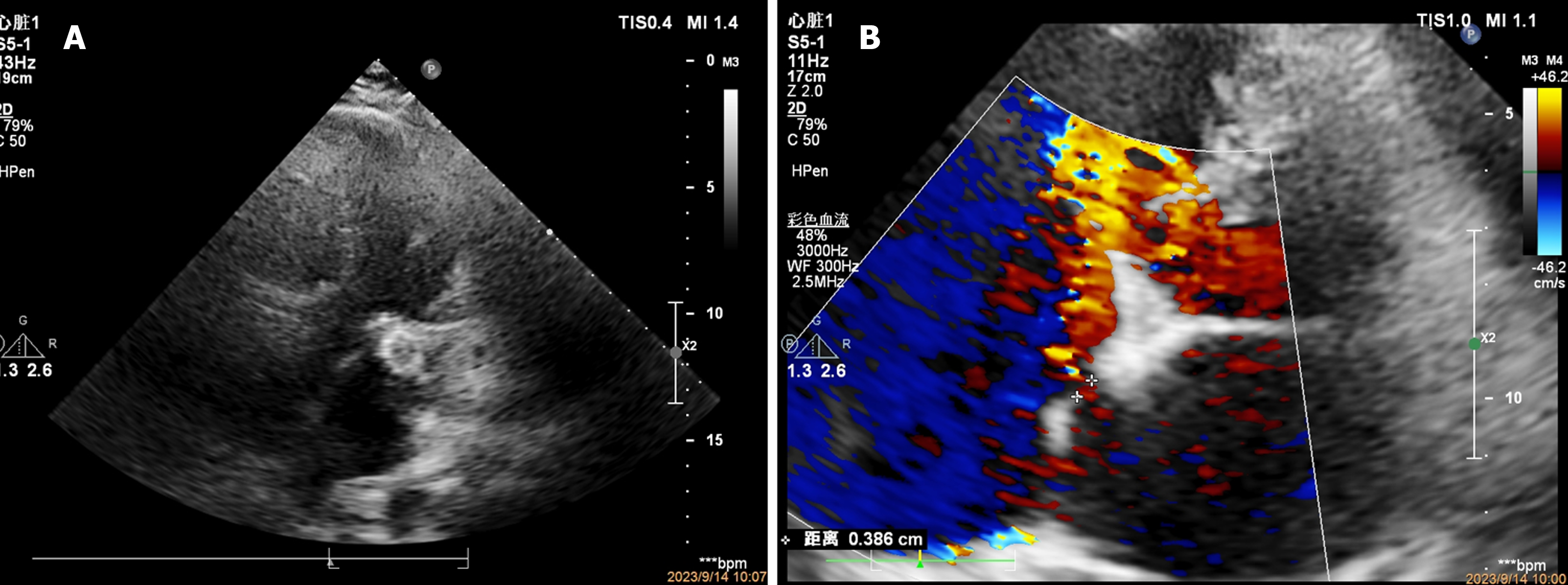
Figure 4 Transthoracic echocardiography.
A: Transthoracic echocardiography shows the occluder located at the left atrial appendage opening; B: Transseptal blood flow at the puncture site of the atrial septum.
- Citation: Yu K, Mei YH. Left atrial appendage occluder detachment treated with transthoracic ultrasound combined with digital subtraction angiography guided catcher: A case report. World J Clin Cases 2024; 12(6): 1157-1162
- URL: https://www.wjgnet.com/2307-8960/full/v12/i6/1157.htm
- DOI: https://dx.doi.org/10.12998/wjcc.v12.i6.1157


