Copyright
©The Author(s) 2024.
World J Clin Cases. Feb 16, 2024; 12(5): 1029-1032
Published online Feb 16, 2024. doi: 10.12998/wjcc.v12.i5.1029
Published online Feb 16, 2024. doi: 10.12998/wjcc.v12.i5.1029
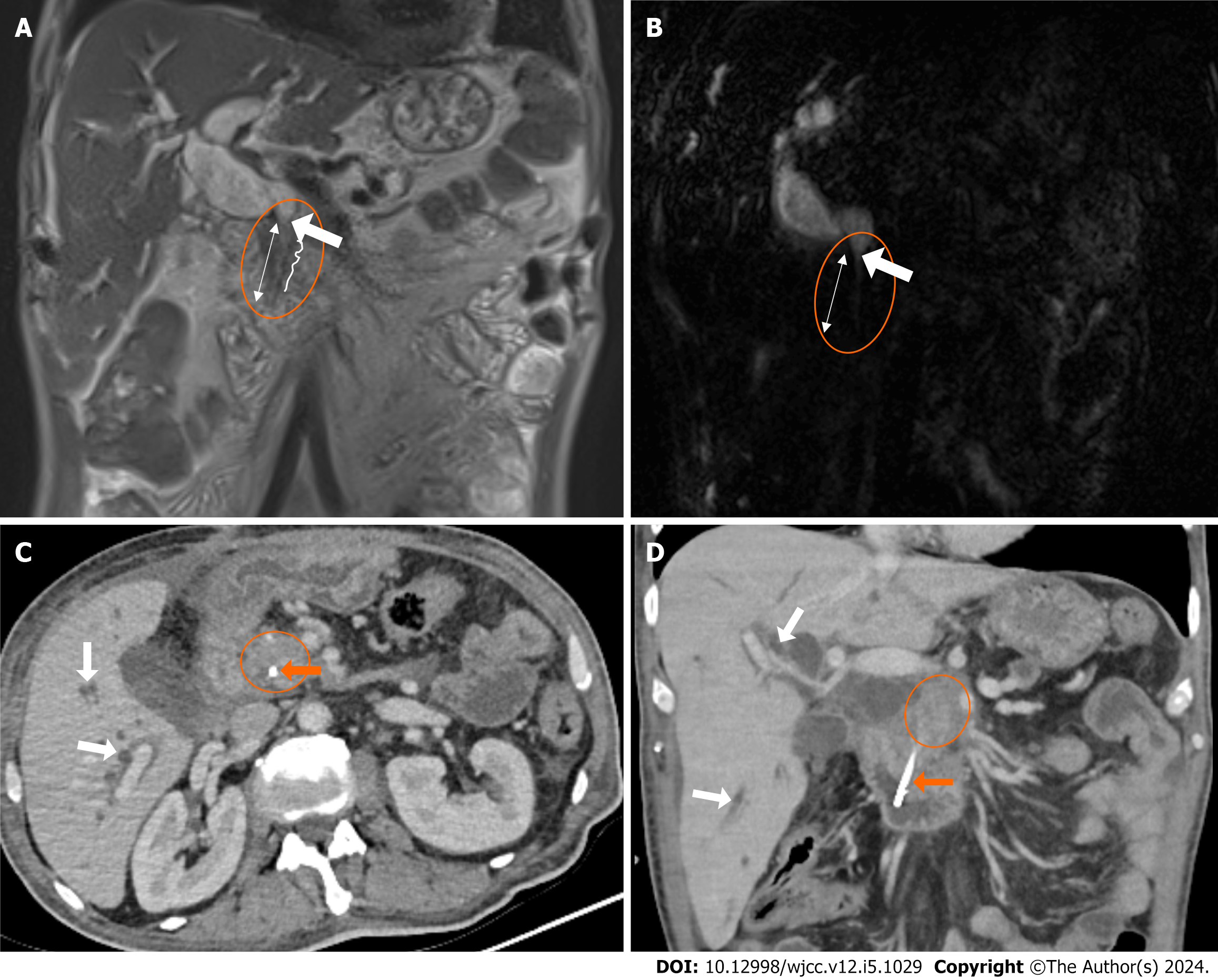
Figure 2 Choledochal involvement of a pancreatic mass.
A and B: Coronal T2 WI and coronal magnetic resonance cholangiopancreatography images. The dilated choledochal duct (circle) abruptly narrows bluntly (white arrow) and continues narrowly in a long segment more distally (two-headed arrow). Contour irregularities (serrated lines) are seen on the distal walls of the choledochal duct; C and D: Post-treatment axial and coronal computed tomography images of the same patient show an irregularly bordered, hypodense, heterogeneous, solid mass lesion (circle) in the head of the pancreas, stent material extending from the duodenum to the pancreas (orange arrow), and dilated intrahepatic bile ducts (white arrow).
- Citation: Aydin S, Irgul B. Response letter to “Acute cholangitis: Does malignant biliary obstruction vs choledocholithiasis etiology change the outcomes?” with imaging aspects. World J Clin Cases 2024; 12(5): 1029-1032
- URL: https://www.wjgnet.com/2307-8960/full/v12/i5/1029.htm
- DOI: https://dx.doi.org/10.12998/wjcc.v12.i5.1029


