Copyright
©The Author(s) 2024.
World J Clin Cases. Feb 6, 2024; 12(4): 828-834
Published online Feb 6, 2024. doi: 10.12998/wjcc.v12.i4.828
Published online Feb 6, 2024. doi: 10.12998/wjcc.v12.i4.828
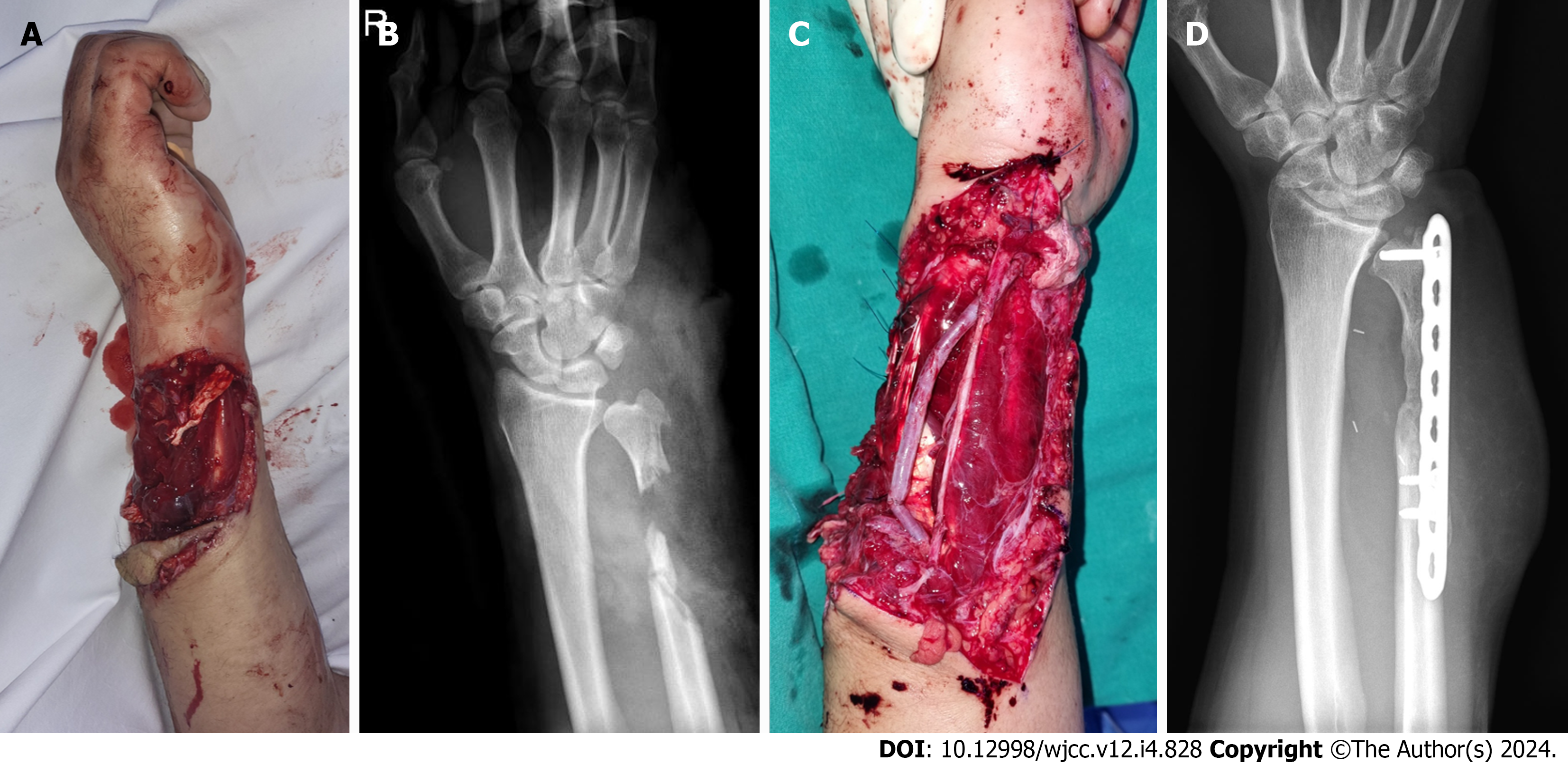
Figure 1 Initial clinical photographs and plain radiographs of the right forearm.
A and B: The 15 cm × 10 cm soft tissue defect of the forearm and open fracture with bone defect of distal ulnar bone were found; C and D: Initially, the ulnar nerve, artery, and tendons were reconstructed, and internal fixation with bone cement were done using the Masquelet technique.
- Citation: Jeon JH, Kim KW, Jeon HB. Pedicled abdominal flap using deep inferior epigastric artery perforators for forearm reconstruction: A case report. World J Clin Cases 2024; 12(4): 828-834
- URL: https://www.wjgnet.com/2307-8960/full/v12/i4/828.htm
- DOI: https://dx.doi.org/10.12998/wjcc.v12.i4.828


