Copyright
©The Author(s) 2024.
World J Clin Cases. Dec 26, 2024; 12(36): 6926-6934
Published online Dec 26, 2024. doi: 10.12998/wjcc.v12.i36.6926
Published online Dec 26, 2024. doi: 10.12998/wjcc.v12.i36.6926
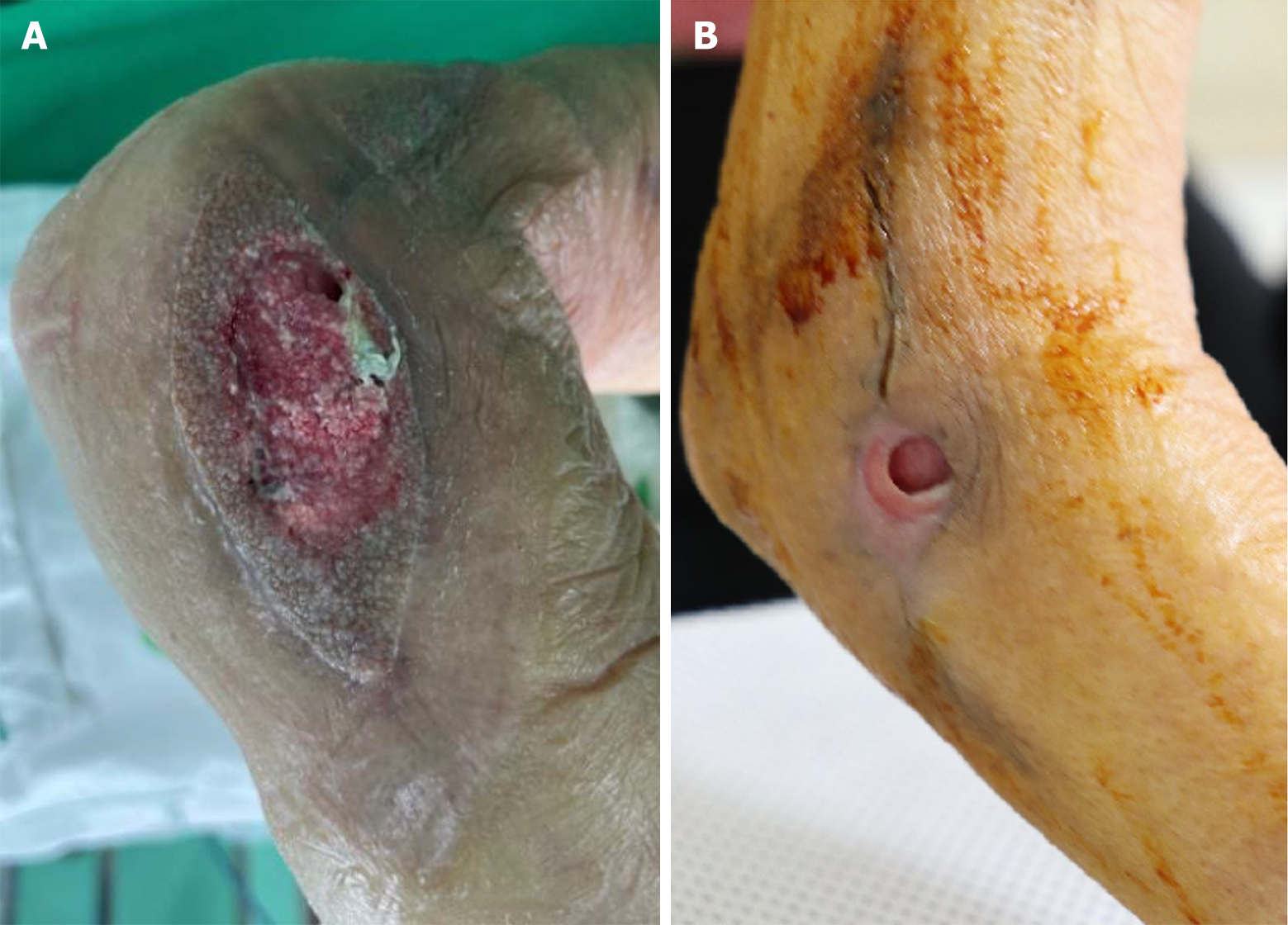
Figure 1 Clinical photographs of the right elbow joint.
A: At treatment onset, the defect (approximately 5.0 cm × 3.0 cm) showed unhealthy granulation tissues, including a partially necrotic change with bone exposure; B: After 4 months of treatment, the dimension of the wound and the amount of exudate were gradually reduced, but the wound stalled without further improvement.
- Citation: Kim JH, Koh IC, Lim SY, Kang SH, Kim H. Chronic intractable nontuberculous mycobacterial-infected wound after acupuncture therapy in the elbow joint: A case report. World J Clin Cases 2024; 12(36): 6926-6934
- URL: https://www.wjgnet.com/2307-8960/full/v12/i36/6926.htm
- DOI: https://dx.doi.org/10.12998/wjcc.v12.i36.6926


