Copyright
©The Author(s) 2024.
World J Clin Cases. Dec 16, 2024; 12(35): 6826-6833
Published online Dec 16, 2024. doi: 10.12998/wjcc.v12.i35.6826
Published online Dec 16, 2024. doi: 10.12998/wjcc.v12.i35.6826
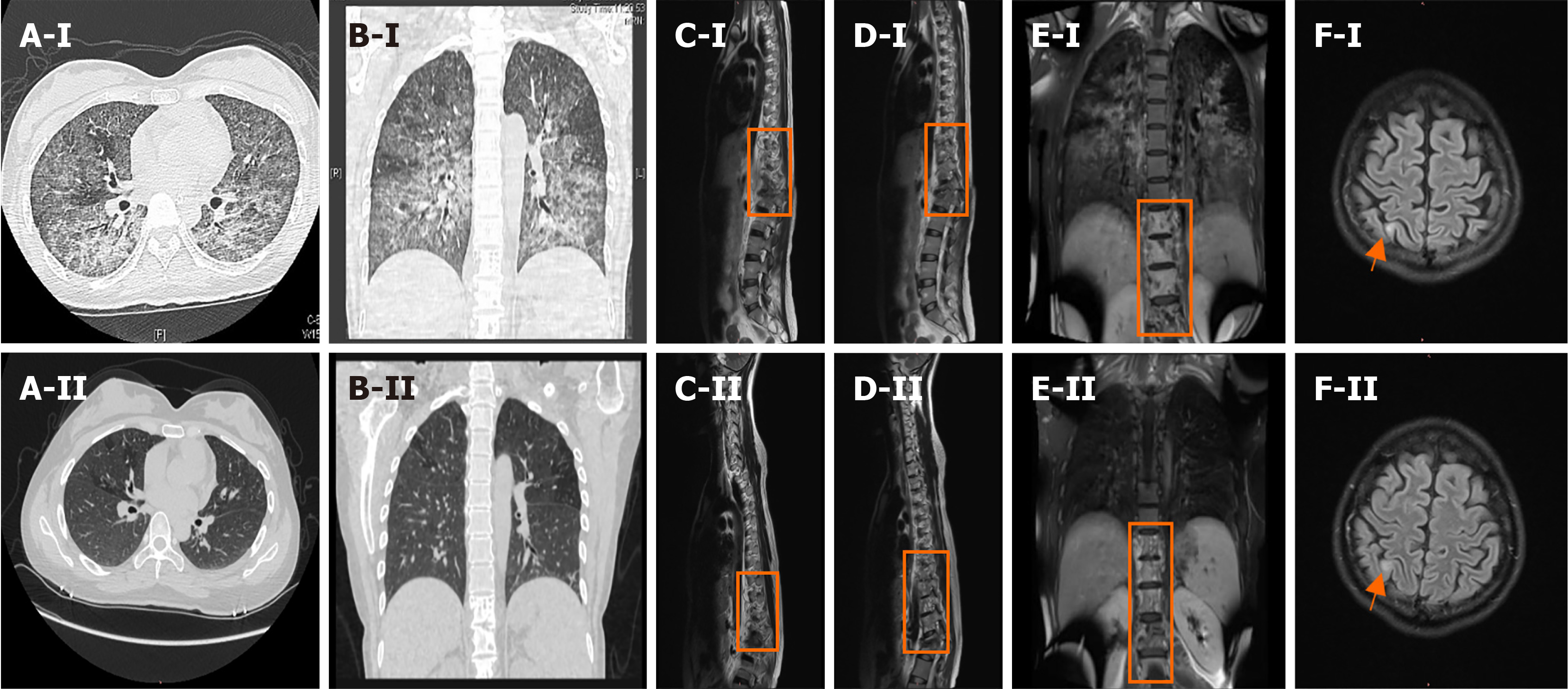
Figure 1 Chest computed tomography and enhanced magnetic resonance imaging of the thoracolumbar spine and brain.
A-I and B-I: Chest computed tomography scans showing diffuse lesions in both lungs on admission; C-I, D-I, and E-I: Enhanced magnetic resonance imaging (MRI) of the thoracolumbar spine performed on day 13, showing abnormal signal shadows from the ninth thoracic vertebral body to the first lumbar vertebral body and the surrounding soft tissues; F-I: Enhanced MRI of the brain performed on day 12, showing an abnormal right parietal lobe signal. A-II and B-II: The chest computed tomography scan performed 6 ½ months after admission, showing an improvement in the diffuse lesions in both lungs. C-II, D-II, and E-II: Enhanced MRI of the thoracic spine performed 6 ½ months after admission, showing improved signal shadows from the ninth thoracic vertebral body to the first lumbar vertebral body and the surrounding soft tissues; F-II: Enhanced MRI of the brain performed 6 ½ months after admission, showing similar signal intensities in the right parietal lobe and the frontal lobe.
- Citation: Wu F, Yang B, Xiao Y, Ren LL, Chen HY, Hu XL, Pan YY, Chen YS, Li HR. psk1 virulence gene-induced pulmonary and systemic tuberculosis in a young woman with normal immune function: A case report. World J Clin Cases 2024; 12(35): 6826-6833
- URL: https://www.wjgnet.com/2307-8960/full/v12/i35/6826.htm
- DOI: https://dx.doi.org/10.12998/wjcc.v12.i35.6826


