Copyright
©The Author(s) 2024.
World J Clin Cases. Nov 6, 2024; 12(31): 6506-6512
Published online Nov 6, 2024. doi: 10.12998/wjcc.v12.i31.6506
Published online Nov 6, 2024. doi: 10.12998/wjcc.v12.i31.6506
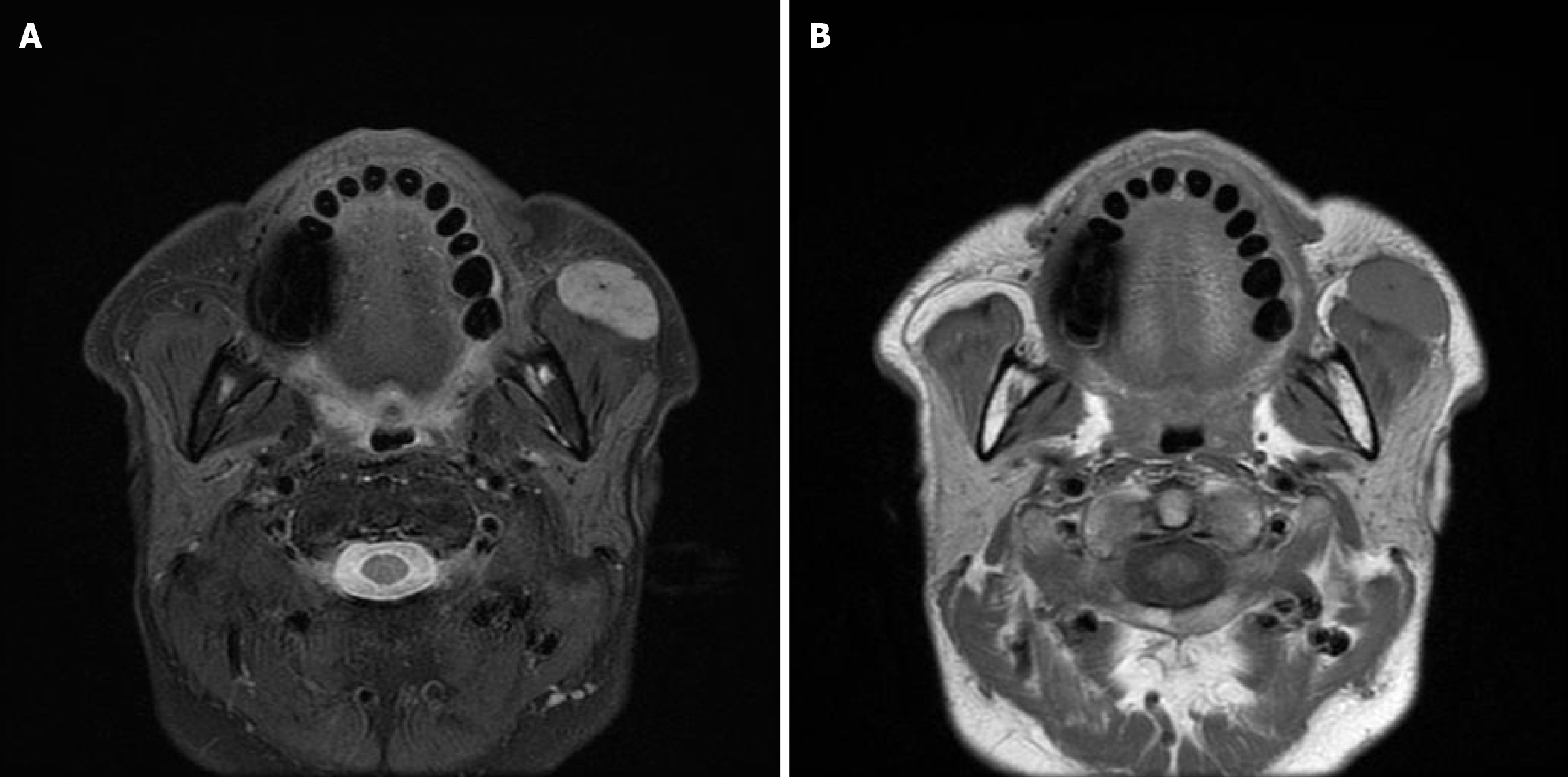
Figure 2 Magnetic resonance images.
A: T1-weighted magnetic resonance images; B: T2-weighted magnetic resonance images. A 30-mm large mass with well-defined margins was observed in the buccal fat body anterior to the left masseter muscle; both T1 and T2 are nonspecific, with moderate signals. No continuity with the parotid glands is noted.
- Citation: Miyake K, Hirasawa K, Nishimura H, Tsukahara K. Rare incidence of mucosa-associated lymphoid tissue lymphoma presenting as buccal fat pad tumor: A case report. World J Clin Cases 2024; 12(31): 6506-6512
- URL: https://www.wjgnet.com/2307-8960/full/v12/i31/6506.htm
- DOI: https://dx.doi.org/10.12998/wjcc.v12.i31.6506


