Copyright
©The Author(s) 2024.
World J Clin Cases. Nov 6, 2024; 12(31): 6486-6492
Published online Nov 6, 2024. doi: 10.12998/wjcc.v12.i31.6486
Published online Nov 6, 2024. doi: 10.12998/wjcc.v12.i31.6486
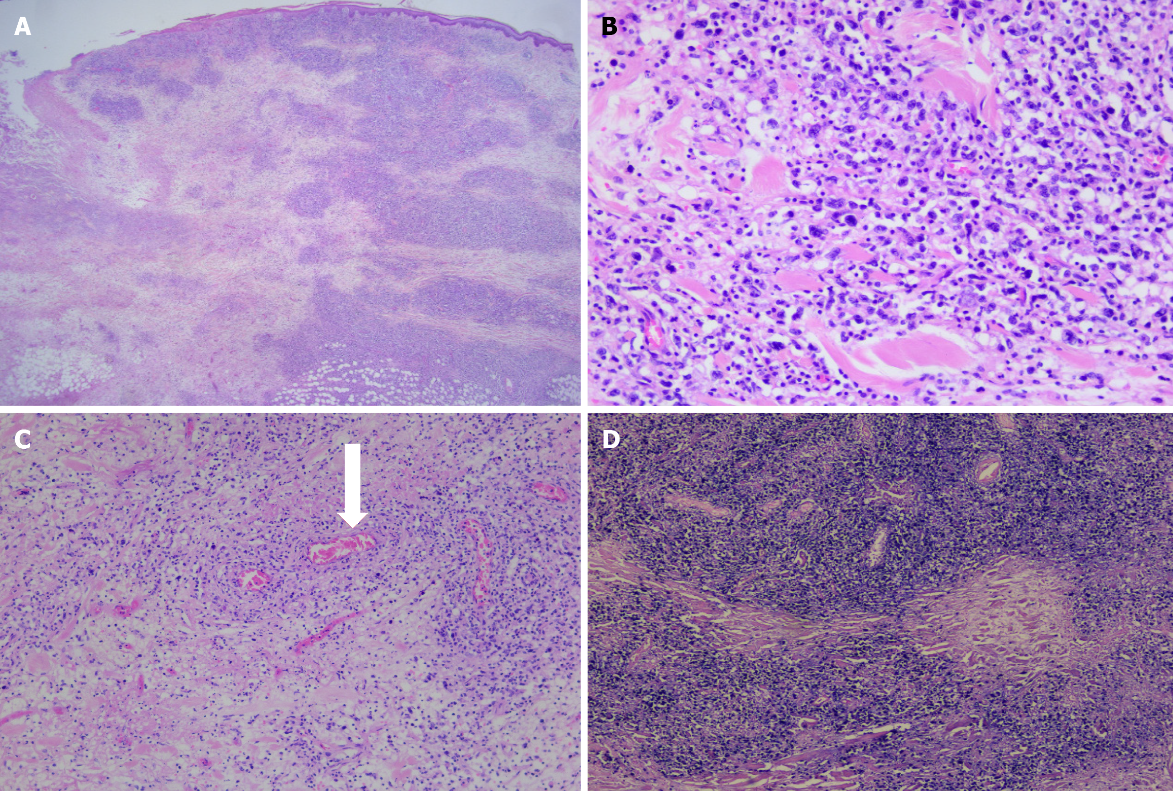
Figure 3 Morphologic and vascular features of tumor necrosis.
A and B: Microscopically, in the background of multifocal geographic tumor necrosis, highly pleomorphic lymphoid cell infiltration is noted; C: Vascular invasion with surrounding necrosis is identified and marked with white arrow; D: In situ hybridization for Epstein–Barr virus-encoded RNA (EBER) showing reactivity (A: H&E, × 5; B: H&E, × 20; C: H&E, × 10; D: EBER in situ hybridization, × 20).
- Citation: Kim JM, Choi WY, Cheon JS. Diagnostic and management challenges in primary cutaneous anaplastic large cell lymphoma with necrosis, inflammation, and surgical intervention: A case report. World J Clin Cases 2024; 12(31): 6486-6492
- URL: https://www.wjgnet.com/2307-8960/full/v12/i31/6486.htm
- DOI: https://dx.doi.org/10.12998/wjcc.v12.i31.6486


